Team:DTU-Denmark/Protein Models
From 2013.igem.org
| Line 9: | Line 9: | ||
Figure 1: Ammonia monooxygenase. Homology models of all three subunits of AMO. | Figure 1: Ammonia monooxygenase. Homology models of all three subunits of AMO. | ||
| - | At the time of writing | + | At the time of writing none of the three subunits from ''Nitrosomonas Europaea'' are available as 3D structures, so each of the subunits was identified at [http://www.uniprot.org/ UNIPROT], and their sequence was homology modelled using [http://www.cbs.dtu.dk/services/CPHmodels/ CPHmodels-3.2] |
{| class="wikitable" | {| class="wikitable" | ||
Revision as of 14:05, 3 October 2013
Protein Models
Contents |
We have determined the structures of some proteins we want our mutants to express. Others could be homology modelled due to existing 3D models that had high similarity to the ones we are working with.
Mutant 1
AMO
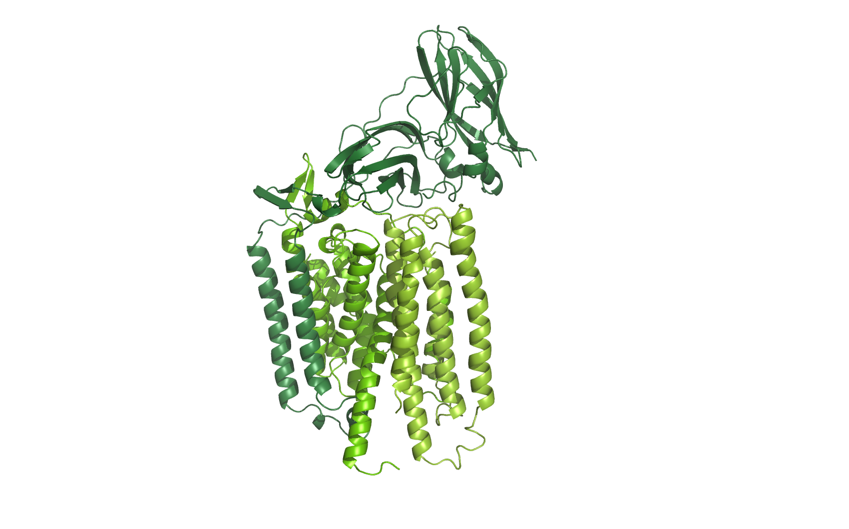
Figure 1: Ammonia monooxygenase. Homology models of all three subunits of AMO.
At the time of writing none of the three subunits from Nitrosomonas Europaea are available as 3D structures, so each of the subunits was identified at [http://www.uniprot.org/ UNIPROT], and their sequence was homology modelled using [http://www.cbs.dtu.dk/services/CPHmodels/ CPHmodels-3.2]
| Resource | AmoA | AmoB | AmoC |
|---|---|---|---|
| Uniprot | [http://www.uniprot.org/uniprot/Q04507 Q04507] | [http://www.uniprot.org/uniprot/Q04508 Q04508] | [http://www.uniprot.org/uniprot/Q82T63 Q82T63] |
| Template pdb code and chain | [http://www.pdb.org/pdb/explore/explore.do?structureId=1yew 1YEW.A] | [http://www.pdb.org/pdb/explore/explore.do?structureId=1yew 1YEW.B] | [http://www.pdb.org/pdb/explore/explore.do?structureId=3rgb 3RGB.C] |
| Coverage | 90.5 | 86.2 | 66.4 |
| E-value | 1e-87 | 3e-67 | 8e-43 |
The three subunits were combined and aligned to the subunits of the [http://www.pdb.org/pdb/explore/explore.do?structureId=1yew Crystal structure] of particulate methane monooxygenase, hence an overlap of two alpha helices.
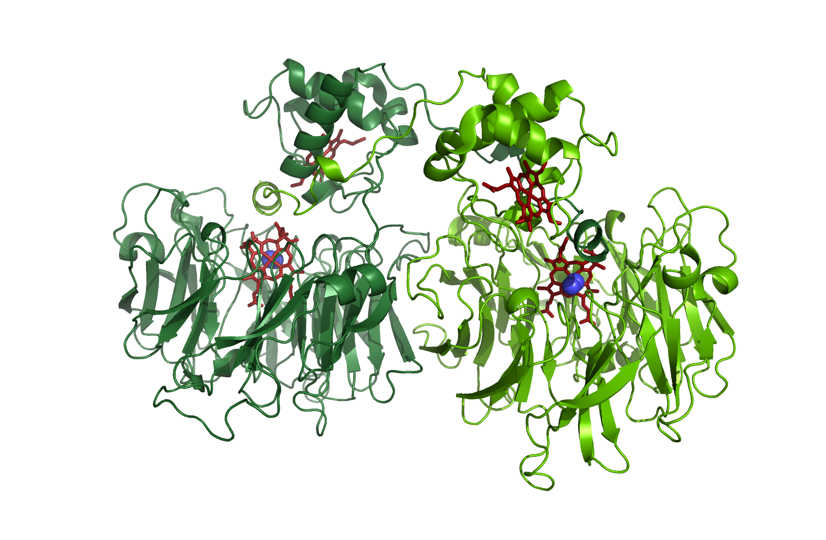
HAO
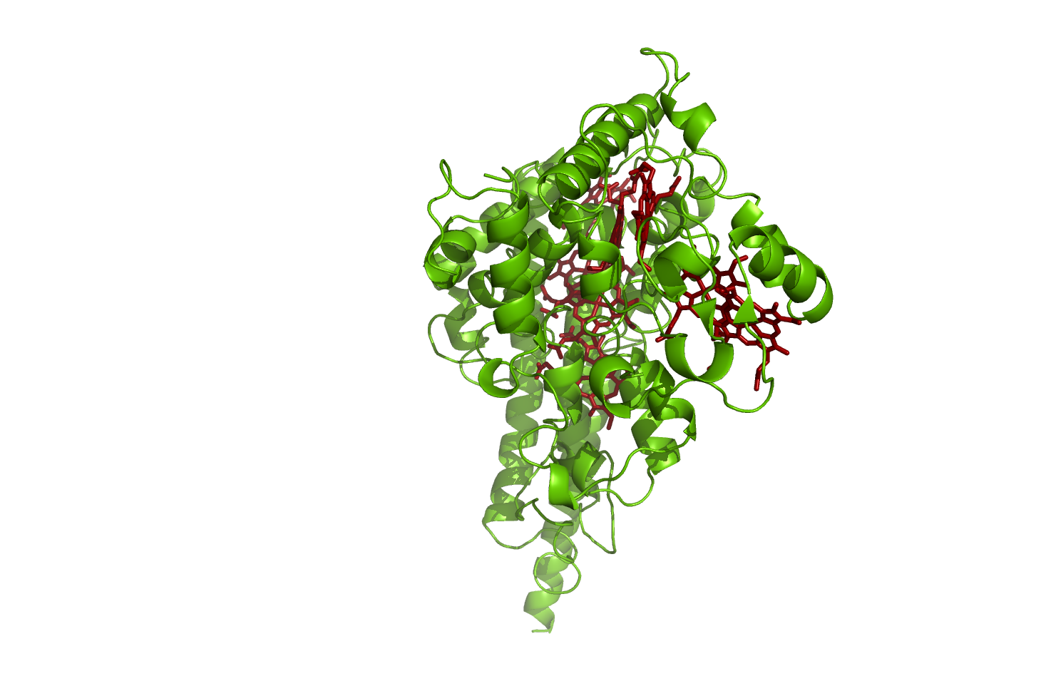
Figure 2: Hydroxylamine oxidoreductase.
source: [http://www.rcsb.org/pdb/explore.do?structureId=1fgj 1FGJ]
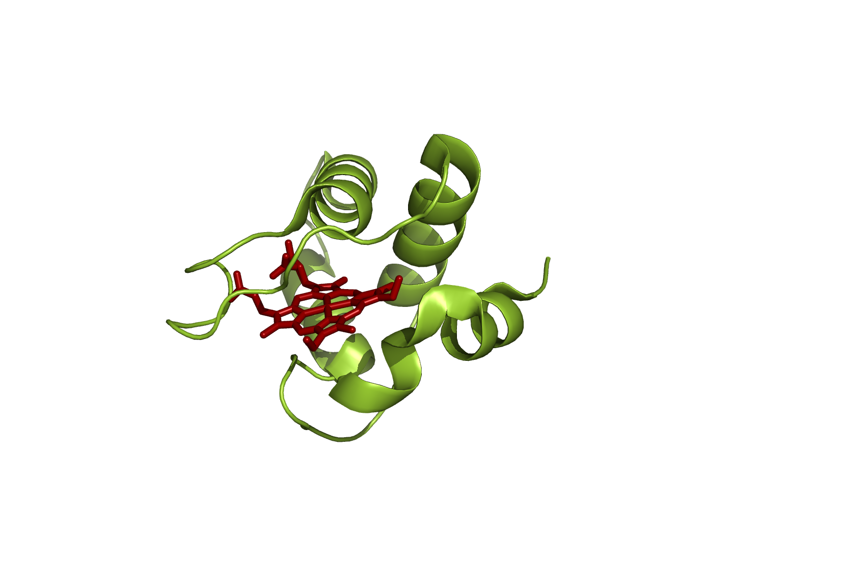
Cc554
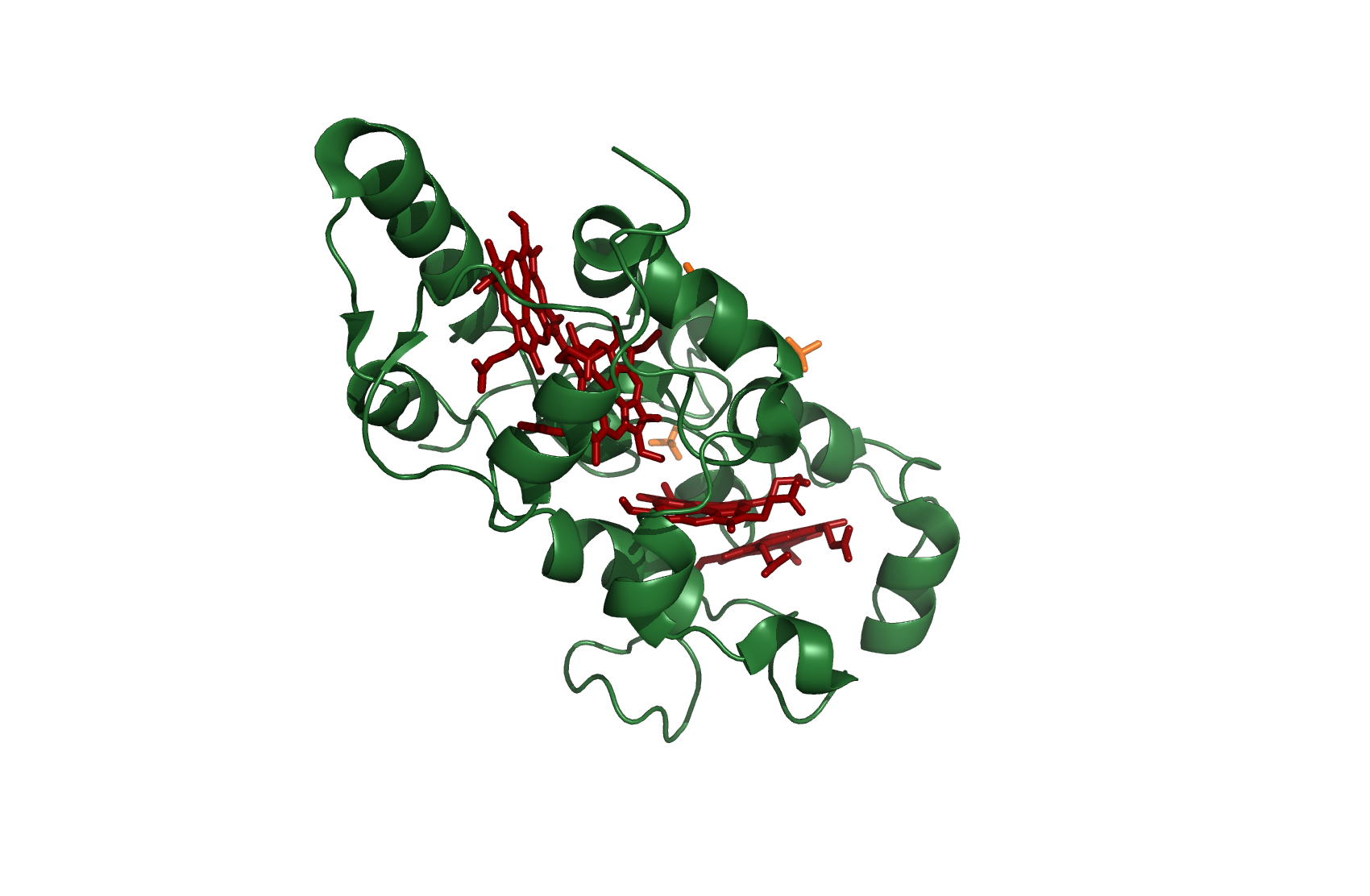
Figure 2: Cytochrome c554
source [http://www.rcsb.org/pdb/explore.do?structureId=1ft5 1FT5]
Ccm552
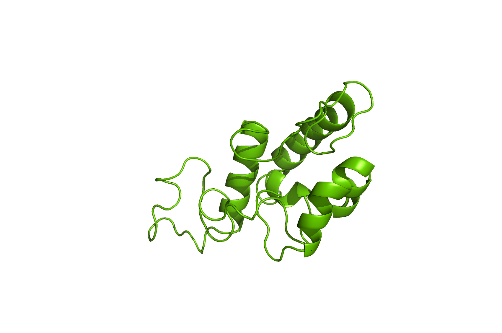
Homology model of Ccm552
| Resource | Ccm552 |
|---|---|
| Uniprot | [http://www.uniprot.org/uniprot/Q50926 Q50926] |
| Template pdb code and chain | [http://www.pdb.org/pdb/explore/explore.do?structureId=1j7a 2J7A.O] |
| Coverage | 68.3 |
| E-value | 6e-11 |
Mutant 2
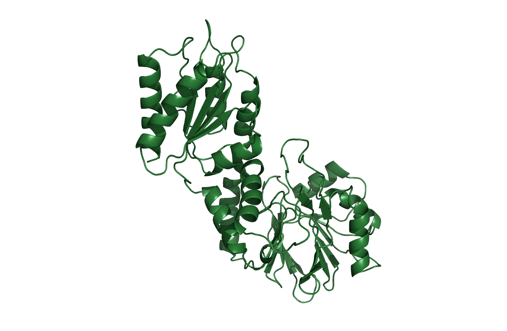
NirS
Nitrite reductase
As a dimer with N2 - source [http://www.rcsb.org/pdb/explore/explore.do?structureId=1nno 1NNO]
NirM
Cytochrom c551
source [http://www.rcsb.org/pdb/explore/explore.do?structureId=2exv 2EXV]
NOR
Flavorubredoxin
Homology model of NOR
| Resource | NOR |
|---|---|
| Uniprot | [http://www.uniprot.org/uniprot/Q46877 Q46877] |
| Template pdb code and chain | [http://www.pdb.org/pdb/explore/explore.do?structureId=1ycf 1YCF.A] |
| Coverage | 82.5 |
| E-value | 3e-95 |
- All homology models were made using the free online tool [http://www.cbs.dtu.dk/services/CPHmodels/ CPHmodels v3.2]
- All models were made and visualized in [http://sourceforge.net/projects/pymol/ PyMOL v1.6] which was easily compiled and installed using an [http://pymolwiki.org/index.php/User:Tlinnet/Linux_Install installation script] made by Troels Linnet.
 "
"
