Team:Heidelberg/Template/Del week18 Gibson
From 2013.igem.org
(Difference between revisions)
(Created page with "==30-08-2013== ===SDS-PAGE=== ====Lysis of cells==== * 1 ml over night cultures in LB of electrocompetent DH10ß, DH10ß with pSB4K5 and DH10ß with pFSN * meausured ODs (DH10ß=...") |
|||
| (2 intermediate revisions not shown) | |||
| Line 1: | Line 1: | ||
| + | |||
==30-08-2013== | ==30-08-2013== | ||
===SDS-PAGE=== | ===SDS-PAGE=== | ||
| Line 18: | Line 19: | ||
'''Results:''' | '''Results:''' | ||
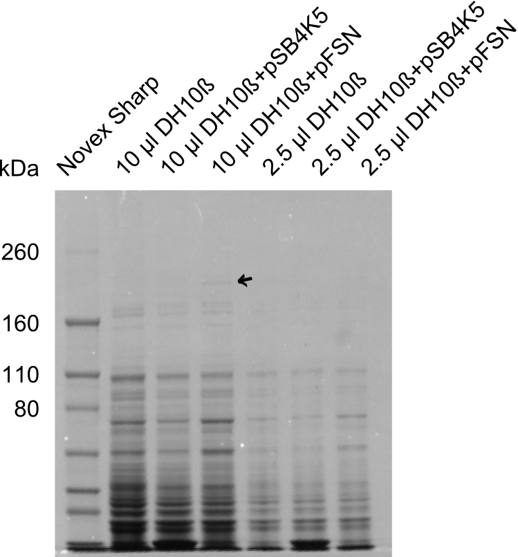
| - | [[File: | + | [[File:Heidelberg_Del1besch.png |150px|thumb | SDS-PAGE of the samples DH10β, DH10β+pSB4K5 and DH10β+pFSN; arrow indicates band of DelE]] |
* One band at a height of about 190 kDa was visible in DH10ß+pFSN, but not in the controls. As the protein DelE has a molecular weight of 192.52 kDa this band is probably DelE. | * One band at a height of about 190 kDa was visible in DH10ß+pFSN, but not in the controls. As the protein DelE has a molecular weight of 192.52 kDa this band is probably DelE. | ||
* A very weak band could be detected well above the 260 kDa band of the ladder. We suspect that this might be DelG, as it has a molecular weight of 357.33 kDa, however the band was too weak to capture it in a picture. Thus a bigger amount of the samples have to be applied on a SDS-PAGE again. | * A very weak band could be detected well above the 260 kDa band of the ladder. We suspect that this might be DelG, as it has a molecular weight of 357.33 kDa, however the band was too weak to capture it in a picture. Thus a bigger amount of the samples have to be applied on a SDS-PAGE again. | ||
<div style="clear:both"></div> | <div style="clear:both"></div> | ||
Latest revision as of 12:44, 3 October 2013
Contents |
30-08-2013
SDS-PAGE
Lysis of cells
- 1 ml over night cultures in LB of electrocompetent DH10ß, DH10ß with pSB4K5 and DH10ß with pFSN
- meausured ODs (DH10ß= 1,32; DH10ß+pSB4K5= 1,28; DH10ß+pFSN= 1,12)
- centrifugation at 13000 rpm for 10 min
- discarded supernatant
- resuspended in 66 µl(DH10ß), 64 µl(DH10ß+pSB4K5) and DH10ß+pFSN= 56 µl 1x loading buffer (40% ddH2O, 12.5% TrisHCl pH=7.5, 20% Glycerol (50%), 20% SDS (10%), 5% β-Mercaptoethanol, 2.5% Bromophenol blue)
- boiled samples for 10 min at 98°C
Electrophoresis
- Applied 2.5 µl and 10 µl of each sample on SDS-polyacrylamide gel
- Let it run for 1 h at 180 V
Staining and destaining procedure
- Gel was stained in Coomassie Brilliant Blue for 1 h on shaker
- Destaining with destaining buffer (50% ddH2O, 40% Methanol, 10% Acetic acid) until bands were visible
Results:
- One band at a height of about 190 kDa was visible in DH10ß+pFSN, but not in the controls. As the protein DelE has a molecular weight of 192.52 kDa this band is probably DelE.
- A very weak band could be detected well above the 260 kDa band of the ladder. We suspect that this might be DelG, as it has a molecular weight of 357.33 kDa, however the band was too weak to capture it in a picture. Thus a bigger amount of the samples have to be applied on a SDS-PAGE again.
 "
"
