Team:Clemson/Notebook

From 2013.igem.org

(Difference between revisions)
Line 13: Line 13:
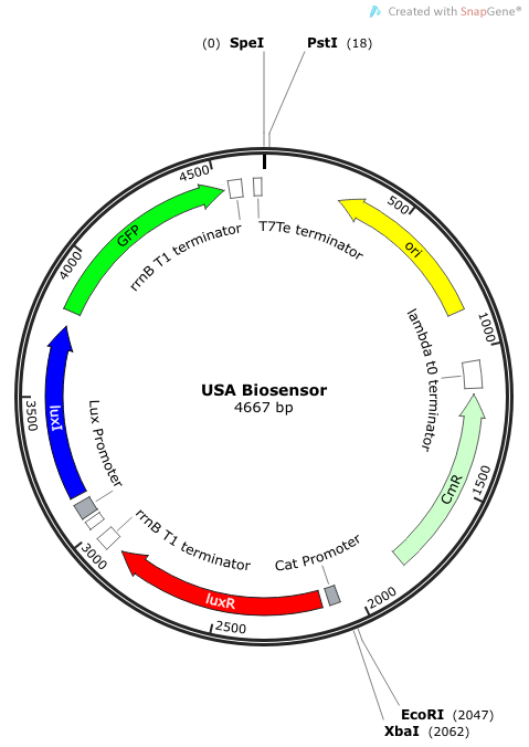
The plasmid construct for the USAB-GFP version is shown below. The other version would replace GFP with amilCP or mAAA.
|-   
|-   
-
|[[File:USA Biosensor.png|175px|frameless|center]]
+
|[[File:USA Biosensor.png|250px|frameless|center]]
-
|[[File:CUTable1.png|175px|frameless|center]]
+
|[[File:CUTable1.png|400px|frameless|center]]
 +
|-
<br>  
<br>  
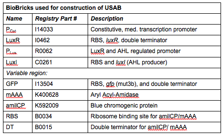
''Rationale'': I14033 was used because the medium transcription rate would be less energetically burdensome on the cell, and this seemed reasonable for a transcriptional regulator. The purpose of having I0462, R0062, and C0261 was to create a positive feedback loop.  I0462 would be activated by an outside source of AHLs thereby activating R0062.  R0062 would then drive transcription of C0261 to express LuxI, which is responsible for synthesizing more AHLs to drive I0462.  This positive feedback loop is what makes the sensor self-amplifying, thus making a very minute signal from a pathogen become more detectable in smaller concentrations.  Detection is made possible by GFP or the colorimetric proteins amilCP and mAAA.

Revision as of 22:12, 27 September 2013

Notebook

Our goal for this year’s project was to construct a universal self-amplifying biosensor (USAB) which could be used to quickly determine the presence or absence of any bacterial pathogen. The self-amplifying nature of our biosensor would allow for rapid and sensitive detection, and the universality of our biosensor would allow it to be adapted to detection of any bacterium. For more information on how our system would work, please see the Project page.
We wanted our system to respond to and amplify the quorum sensing AHL signals that would be sent from a pathogen (more details on this later), and respond by sending out a visible signal that we could measure or detect (e.g. GFP, chromogenic protein, etc.). An outline of our approach to synthesize and test our USAB is detailed below.

Phase I: Construction of the Universal Self-Amplifying BioSensor (USAB) We actually decided to make three version of this; each would include the constitutively expressed activator luxR, the LuxR/AHL activated promoter PLux followed by the AHL-producing luxI and a signal output (GFP, amilCP, or mAAA). GFP would, of course, produce a fluorescent signal, amilCP is a blue chromogenic protein, or mAAA would enzymatically convert the colorless acetomenaphin to a reddish compound. The goal was to find out which of the three versions could inherently produce the most easily detectable signal output in the shortest amount of time.
The plasmid construct for the USAB-GFP version is shown below. The other version would replace GFP with amilCP or mAAA.
Rationale: I14033 was used because the medium transcription rate would be less energetically burdensome on the cell, and this seemed reasonable for a transcriptional regulator. The purpose of having I0462, R0062, and C0261 was to create a positive feedback loop. I0462 would be activated by an outside source of AHLs thereby activating R0062. R0062 would then drive transcription of C0261 to express LuxI, which is responsible for synthesizing more AHLs to drive I0462. This positive feedback loop is what makes the sensor self-amplifying, thus making a very minute signal from a pathogen become more detectable in smaller concentrations. Detection is made possible by GFP or the colorimetric proteins amilCP and mAAA.
Note: We also constructed prototype operons composed of the BioBrick parts R0010 (lac promoter) and I13504 (RBS-GFP-DT), or B0034-K400628-B0015 (mAAA version), or B0034-K592009-B0015 (amilCP version). We qualitatively determined that ligation of R0010 and I13504 and the subsequent transformation of R0010 + I13504 into E. coli was functional due to green florescence under UV light. We were never able to see blue color formation from amilCP or red color formation from mAAA. There may have been a mistake in our process for amilCP (e,g, missing RBS perhaps) to account for our results. See our “Experience” page for mAAA for more info on that.

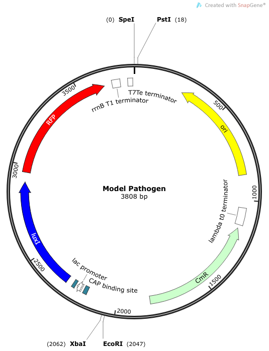
Phase II: Construction of “Model Pathogen” We needed a bacterium that would produce an initial AHL signal to stimulate the USAB. Eventually a similar construct would be delivered via phage to specific pathogens, but for now we needed an easy way to test the self-amplifiying ability of our USAB.
USA Biosensor.png
CUTable1.png
Model Pathogen.png