Team:Carnegie Mellon/Parts
From 2013.igem.org
| Line 2: | Line 2: | ||
<br><br> | <br><br> | ||
<h1>Parts</h1> | <h1>Parts</h1> | ||
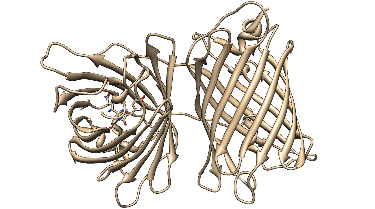
| - | [[image:KillerRed.png| | + | [[image:KillerRed.png|400px|thumb|center|Crystal Structure of KillerRed (PDB ID: 3GB3) with the QYG chromophore shown<sup>2,3,4</sup>]] |
<br><br> | <br><br> | ||
Revision as of 06:40, 27 September 2013
Parts
KillerRed (<partinfo>BBa_K1184000</partinfo>) is a red fluorescent protein that produces reactive oxygen species (ROS) in the presence of yellow-orange light (540-585 nm). KillerRed is engineered from anm2CP to be phototoxic. It has been shown that KillerRed produces superoxide radical anions by reacting with water. Superoxide reacts with the chromophore of KillerRed, causing it to become dark, which ultimately gives rise to a bleaching effect. KillerRed is spectrally similar to mRFP1 with a similar brightness. KillerRed is oligomeric and may form large aggregates in cells. Expression of KillerRed and irradiation with light may act a kill-switch for biosafety applications.
The parts we used this year to characterize KillerRed involve the mRFP1 protein (<partinfo>BBa_E1010</partinfo>), which acts as a negative control for phototoxicity due their spectral similarity. For expression, we used a wild-type lac promoter (<partinfo>BBa_R0010</partinfo>) and an RBS (<partinfo>BBa_B0034</partinfo>). mRFP1 was characterized and its experience page was updated with our experimental data. These parts were cloned into a pSB1A2 vector for characterization, however a λ phage was cloned with the KillerRed coding sequence for a demonstration of proof-of-concept.
Bacteriophage Lambda
Esther Lederberg isolated the bacteriophage λ in 1950 while at the University of Wisconsin. The λ that we selected for use is lambda gt11, a vector designed for protein expression and screening by antibodies or nucleic acid hybridization (Young and Davis, 1983). The inserts are cloned into a unique EcoRI site at the C-terminus of the β-galactosidase gene so that fusions destroy the enzymatic activity and allow for a blue-white screen of plaques. There are hosts available that allow for high frequency of lysogenization (HFL).
More Information about KillerRed
More Information about RFP <groupparts>iGEM013 Carnegie_Mellon</groupparts>
References
Lederberg, E. M., 1950, Lysogenicity in Escherichia coli strain K-12, Microbial Genetics Bulletin, 1, pp. 5-9, Jan. 1950, Univ. of Wisconsin (Evelyn Maisel Witkin, Editor), Ohio State University, ISSN: 0026-2579, call No. 33-M-4, OCLC: 04079516, Accession Number: AEH8282UW" http://www.estherlederberg.com/Censorship/LambdaW.html
 "
"

