PET-mCherry
From 2013.igem.org
(→PET-mCherry) |
(→PET-mCherry) |
||
| Line 1: | Line 1: | ||
{{:Team:GeorgiaTech/template}} | {{:Team:GeorgiaTech/template}} | ||
=PET-mCherry= | =PET-mCherry= | ||
| - | [[File:MCherry.PNG|right| | + | [[File:MCherry.PNG|right|200px|border]] |
'''PET-mCherry''' is a β-barrel autotransporter with a mCherry passenger. | '''PET-mCherry''' is a β-barrel autotransporter with a mCherry passenger. | ||
*[http://parts.igem.org/Part:BBa_K1156000# BBa_K1156000] | *[http://parts.igem.org/Part:BBa_K1156000# BBa_K1156000] | ||
Revision as of 00:51, 28 September 2013

Main Page

Contents |
PET-mCherry
PET-mCherry is a β-barrel autotransporter with a mCherry passenger.
- [http://parts.igem.org/Part:BBa_K1156000# BBa_K1156000]
Motivation
In 2012 the Georgia Tech iGEM team developed a novel biosensor based off of green fluorescent protein. The sensor consisted of two subunits of the protein that separately were inactive but once dimerized expressed fluorescence. From this project, we began thinking about how we could develop more complex sensing technology in bacteria. Taking into consideration how mammalian cells sense and react to their environment, we started asking the question: Can bacteria express human integrins?
To start answering this question, we needed to find a way to transport large proteins and anchor them to the outside of the cell. Autodisplay technology seemed like one possible solution to this problem.
Design
In a 2012 paper, a research group from the University of Birmingham demonstrated that the β-barrel PET could successfully transport and translocate the RFP, mCherry.[http://www.microbialcellfactories.com/content/11/1/69|] We wanted to use their construct to determine whether or not PET would be a good candidate for integrin transport. The sequence that was provided in the paper was already optimized for E. coli expression, however we checked it again. There were also a number of compatibility issues that need to be resolved. To comply with BioBrick standard 10, Phe-76 was re-optimized to remove a EcoRI site withing the part. A T7 promoter with a lacI operator was added with the RBS (AGGA) to the PET_mCherry. A standard assembly 10 prefix and suffix was then added. In future work, other autodisplay technologies are hoped to be tested to express a number of proteins.[http://www.uniprot.org/uniprot/O68900 Uniprot]
Because of a generous offer from IDT for discount synthesis we chose to synthesize our protein construct using their gBlock method which consists of blocks of 500bp. Our construct fit into 6 blocks.
Below are the primers that we designed to amplify each block.
Primers:
ig12 SP/Pet_mCherry_b1/0	AGTCAGGAATTCGCGGCCGCTTCTAGAGG
ig13 SP/Pet_mCherry_b2/469	CGTATGAAGGCACCCAGACCGCTAAACTGAAA
ig14 ASP/Pet_mCherry_b1/500	TTTCAGTTTAGCGGTCTGGGTGCCTTCATACG
ig15 SP/Pet_mCherry_b3/928	CGGGTGCTTACAACGTGAACATCAAACTGGAC
ig16 ASP/Pet_mCherry_b2/959	GTCCAGTTTGATGTTCACGTTGTAAGCACCCG
ig17 SP/Pet_mCherry_b4/1352	CAAACTGGAAGGTGCGAACAACCTGCTGC
ig18 ASP/Pet_mCherry_b3/1380	GCAGCAGGTTGTTCGCACCTTCCAGTTTG
ig19 SP/Pet_mCherry_b5/1816	TGTTCACCGGTGTTACCATGACCTACACCGAC
ig20 ASP/Pet_mCherry_b4/1847	GTCGGTGTAGGTCATGGTAACACCGGTGAACA
ig21 SP/Pet_mCherry_b6/2265	GGTTACCAGTTCGACCTGTTCGCTAACGGTGA
ig22 ASP/Pet_mCherry_b5/2296	TCACCGTTAGCGAACAGGTCGAACTGGTAACC
ig23 ASP/Pet_mCherry_b6/2516	ACTCTGCAGCGGCCGCTACTAGTATTATTATC
Assembly
The 6 gBlocks that we synthesized using IDT were assembled using overlap extension PCR.
Characterization
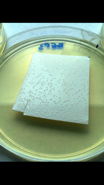
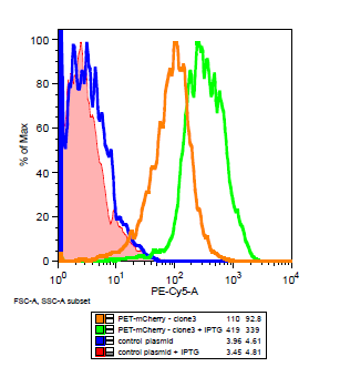
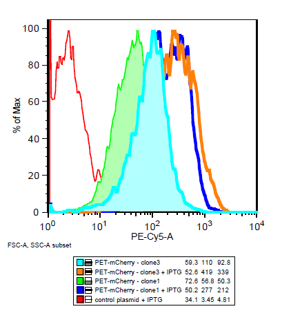
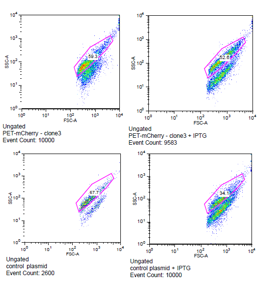
The cells for the colonies BEP1 and BEP3 were characterized using flow cytometry. Our goals for this characterization were to show activity of the LacI operator and confirm the expression of mCherry on the outside of the cell. Cells expressing H6, a single strand antibody that favors fibrin, were used as a negative control.
Is the LacI operator working?
Yes! an increase in mCherry expression after induction with IPTG shows that the LacI operator in conjunction with the T7 promoter is working.
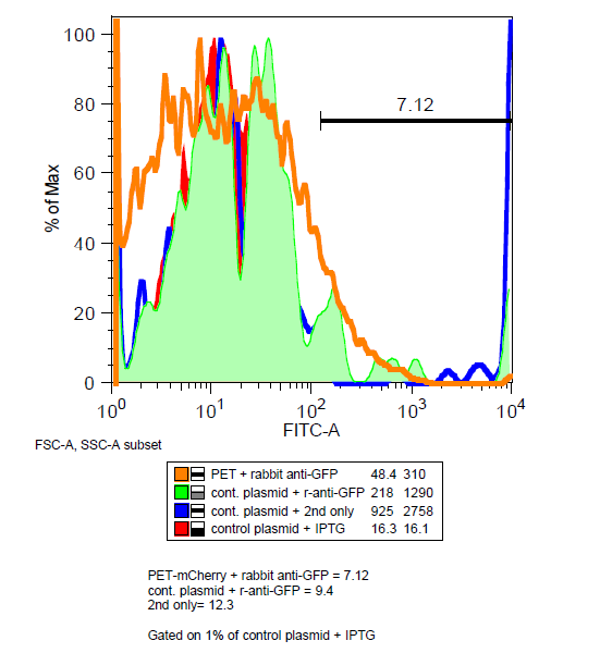
Is mCherry on the outside of the cell?
Maybe... Unlike GFP, mCherry has been shown to be functional in the periplasm.[http://jb.asm.org/content/early/2011/07/15/JB.00315-11.full.pdf] In order to determine if mCherry was being expressed outside the cell, BEP3 cells were stained with mouse and rabbit anti-GFP antibodies and then fluorescent tagged anti-mouse and anti-rabbit antibodies.
Very little decrease in the mCherry signal after washing suggest that the protein is not being cleaved if it is expressed externally. Results of the antibody were inconclusive as the amount of instances for BEP3 was less in the gated data than the control. However a decrease in mCherry signal after treatment of mouse anti-GFP does suggest that at least some of the proteins are on the surface of the cells.
References
[1]Sevastynovich et al."A generalised module for the selective extracellular accumulation of recombinant proteins." Microbial Cell Factories. 2012.
[2]Dinh, T., & Bernhardt, T. G. (2011). Using superfolder green fluorescent protein for periplasmic protein localization studies. Journal of bacteriology, 193(18), 4984-4987.
 "
"
