Team:Heidelberg/Tyrocidine week19 biobrickmod
From 2013.igem.org
Contents |
RFC10 Standardization of Tyc Modules
Ligation of digested Fragments each with linearized pSB1C3
DNA Concentration of Digestions
| Fragment | Source | Protocol | Concentration |
|---|---|---|---|
| Tyc AdCom | AT_01/AT_02 | Reamplification 2013-08-31xxx | 5.0 ng/µl |
| Tyc B1 | AT_09/AT_10 | Reamplification 2013-08-31xxx | 22.5 ng/µl |
| Tyc C5 | AT_11/AT_06 | Reamplification 2013-08-31xxx | 19.2 ng/µl |
| Tyc C6 | AT_07/AT_08 | Reamplification 2013-08-31xxx | 35.7 ng/µl |
| pSB1C3 | mediprep of ONC | - | 14.4 ng/µl |
Reaction
1:1 Ratio of pSB1C3:insert
| Fragment | Size [kb] | Concentration [ng/µL] | Amount for Reaction [µL] | 10x T4 DNA Lig Buffer [µL] | T4 DNA Ligase [µL] | +ddH2O |
|---|---|---|---|---|---|---|
| add Backbone pSB1C3; 4.2 kb; 14.4 ng/µl | 3.5 | |||||
| Tyc AdCom | 3.2 | 5.0 ng/µl | 7.6 | 2 | 1 | 5.9 |
| Tyc B1 | 3.2 | 22.5 ng/µl | 1.7 | 11.8 | ||
| Tyc C5 | 3.2 | 19.2 ng/µl | 2 | 11.5 | ||
| Tyc C6 | 3.9 | 35.7 ng/µl | 1.5 | 12 | ||
Transformation (Standard Protocol) into TOP10
2 plates per Ligation one with less one with more transformed cells.
Picking & Screening I
picked red clones Picked 3 clones per plate= 6 clones per construct
Colony PCR with Amplification Primers
Phusion Flash Colony PCR (see amplification protocols above)
Result NO Product?!
Screening Colony PCR with VR_rv and VF2_fw
iTaq Colony PCR Result NO Product?!
Picking & Screening II
NEGATIVE RESULTS BEFORE BECAUSE: mrfp was replaced by insert thus the positive colonies are WHITE!!! Picked white colonies
Screening PCR
Screened all picked colonies of one fragment: positive Screened one clone per fragment: positive
Tyrocidine-Indigoidine-Fusion - Standardization of Constructs
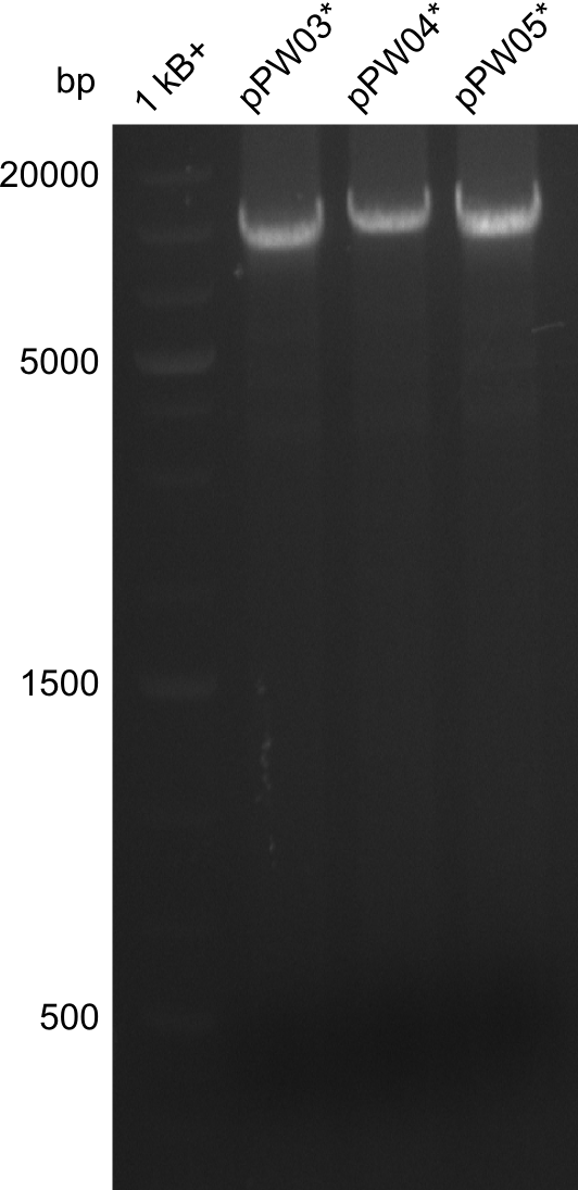
So far, the Tyrocidine-Indigoidine-fusion constructs had two illegal cutting sites of RFC-10 restriction enzymes. Hence, we tried a CPEC-approach for which we reamplified a fragment from the vector with primers that introduced a mutation at the desired position.
Amplification
| what | µl |
|---|---|
| pPW05 (dil.) | 1 |
| RB68 | 2 |
| RB69 | 2 |
| Phusion Flash 2x Master Mix | 10 |
| ddH20 | 5 |
| Cycles | temperature [°C] | Time [min:s] |
|---|---|---|
| 1 | 98 | 0:05 |
| 35 | 98 | 0:05 |
| 59 | 0:10 | |
| 72 | 3:00 | |
| 1 | 72 | 10:00 |
| 1 | 10 | inf |
Results
CPEC
After gel-extraction of the large fragment, the following procedure was followed:
- 22µl H2O, 20µl of the large fragment, 2µl of the short fragment, 1µl DpnI and 5µl 10x CutSmart buffer were incubated for 2 hours at 37°C
- Mixture was purified with isoprop, washed with ethanol and eluted in 10µl H2O
- 10µl Phuson Flash Master Mix was added and the followin protocol was run:
| Cycles | temperature [°C] | Time [min:s] |
|---|---|---|
| 1 | 98 | 0:10 |
| 5 | 98 | 0:01 |
| 53 | 0:05 | |
| 72 | 3:00 | |
| 1 | 72 | 10:00 |
| 1 | 10 | inf |
Unfortunately the efficiency of this was not high enough, as no colonies were visible on the plates after heat-shock transformation in BAP-I cells. The Amplification and CPEC will be repeated next week.
 "
"
