 "
"
Team:BYU Provo/Results/Experimental
From 2013.igem.org
| |
|
|
Phage TeamIsolation of Phage Library
Results of Mutant T4 Phage Isolation

Results of Mutant T7 Phage Isolation As part of creating our bacteriophage library, we wanted to isolate both smaller and larger T7 bacteriophage. To do so, we first mutagenized T7 by growing it in the presence of 5-bromodeoxyuridine, a mutagenic base analog. This produces many mutant T7 bacteriophages, but they are hard to find amongst all the wild type T7. Therefore the phage purification team ran the mutagenized bacteriophage through a cesium chloride gradient. This separates the phage according to size, with the biggest traveling farther down. After determining where the mutant phage is in the gradient, we then plated the phage and looked for plaques that were smaller or larger than normal. Out of the 31 plaques we selected, two of them reproduced their plaque sizes, revealing that they were indeed mutant T7 bacteriophage! This can be seen in the two pictures below. The S4 plate with small phage produced larger plaques, while the L8 plate with large phage produced smaller plaques. The next step will be to get pictures of the phage with an electron microscope and sequence the genome to determine where the mutations occurred. In the two pictures below  
Cholera TeamCholera Induces Bacteriophage Lambda From Lysogeny to its Lytic Cycle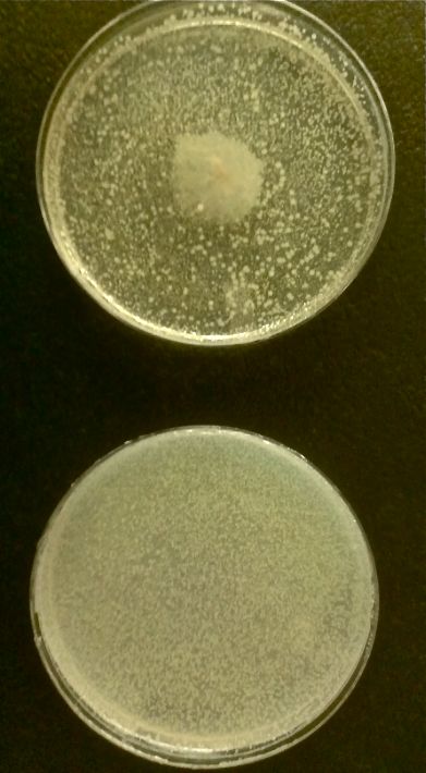
Designing SdiA to be Specific for Cholera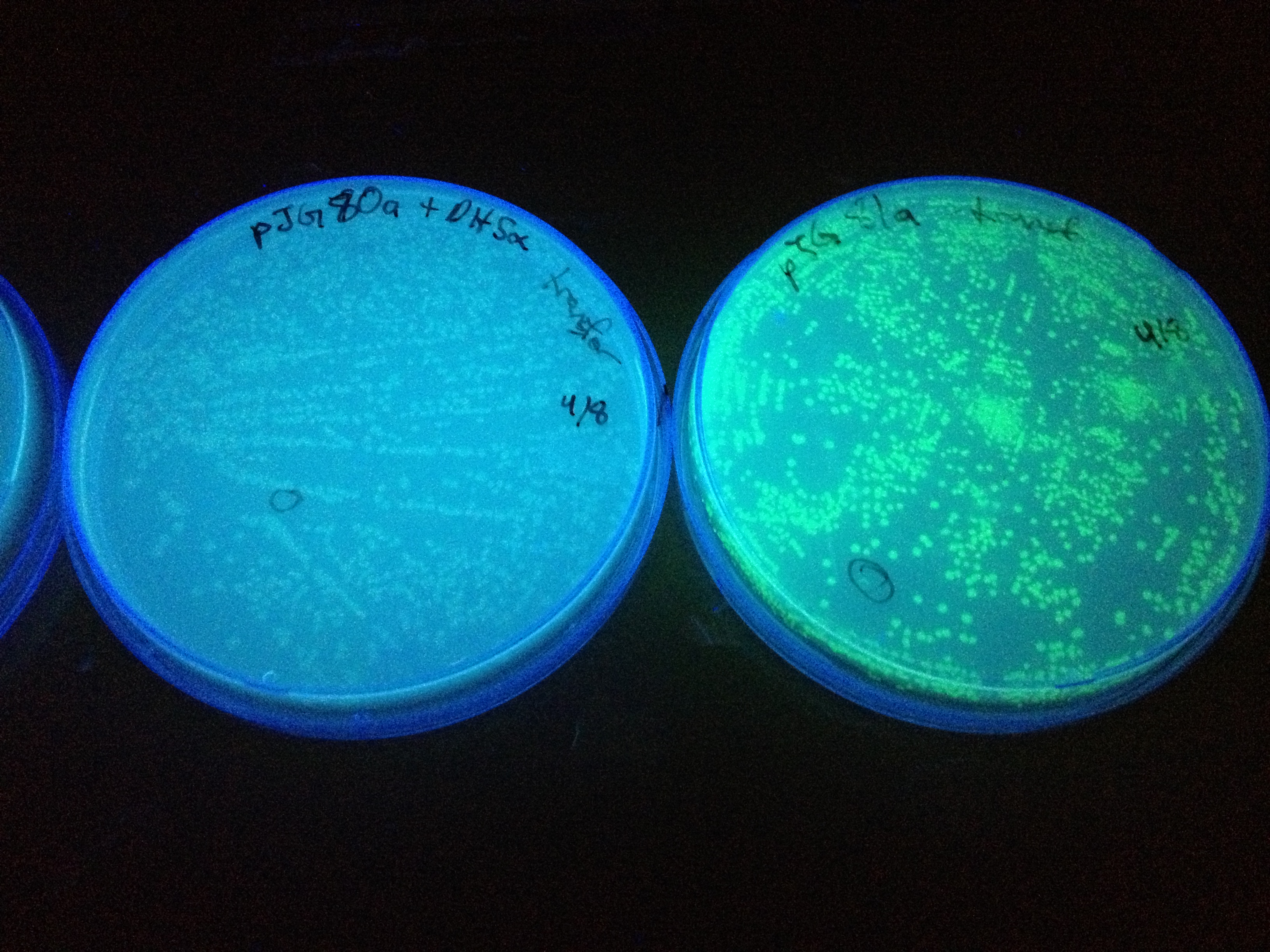 In the beginning our plan was to transform into E.coli the most essential parts of the V.cholerae quorum sensing system. This was done, but our response proteins GFP and RFP did not work as expected.Shown above are colonies that were not supposed to be glowing. So we decided to try mutating the quorum sensing system E.coli already had to be able to only sense CAI-1 which is an auto-inducer specific to V.cholerae. 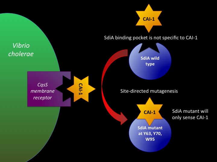 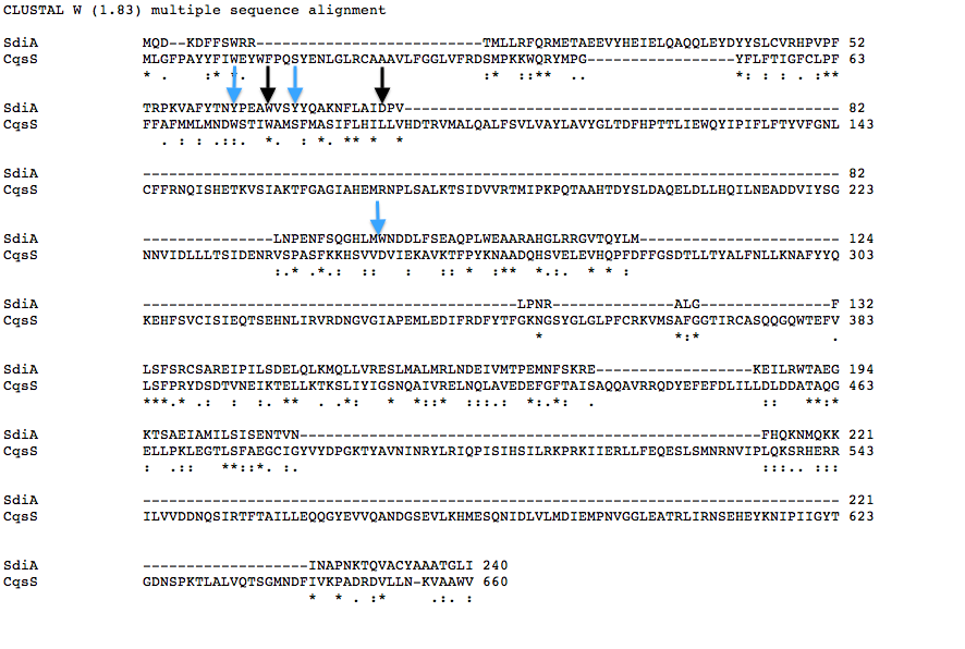
Biofilm inhibition by AmylaseThe enzymatic activity of α-Amylase was characterized to determine its capacity to inhibit biofilm formation by V. cholerae. Samples were prepared by adding 50 µL of V. cholerae culture to 1 mL of a high salt LB. The samples were then treated by the addition of purified α-Amylase in various concentrations and allowed to incubate at 30°C for 48 hours. After 48 hours the samples were examined and a distinct difference was seen in the amount of biofilm formed in the treated and untreated samples. 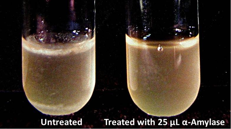 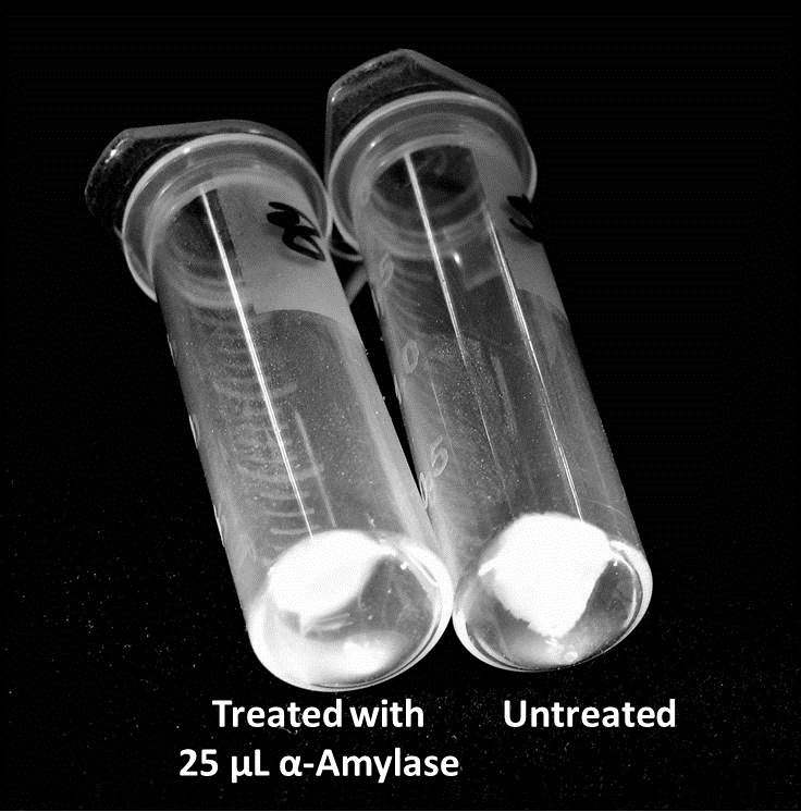
 The above graph shows both the average pellet weight and the average absorbance readings for samples treated with 0, 2.5, 12.5, and 25 µL of α-Amylase. There is a distinct reduction in the amount of biofilm growth between untreated and treated samples, with samples treated with 25 µL α-Amylase showing a 65.8% decrease in biofilm formation after 48 hours. While this clearly shows the ability of α-Amylase to inhibit biofilm formation by V. cholerae, further characterization is needed to determine the capacity of α-Amylase to degrade preexisting biofilms.
|