 "
"
Team:OUC-China/Microfluidics
From 2013.igem.org

Microfluidics
Detecting the magnetism of small amount of bacterial sample is always difficult, because not only the sample is so little that normal magnetic spectrophotometer could not fetch precise data, but also the debugging of magnetic spectrophotometer is quite difficult and expensive for the exist of earth magnetic field.
Since our compartment gene is from Magnetospirillum Magneticum which is able to do directional movement along the geomagnetic field due to its magnetosome, so we need to culture the Magnetospirillum Magneticum AMB-1 strain firstly. While we cultured the AMB-1 bacteria strain, we found that if we hope to evaluate whether our culture scheme is feasible, we should confilm not only the grow, but also the magnetism of the bacteria is normal .So we designed an easy solution of bacteria magnetism detection for smallamount of samplesto make our bacteria magnetism detection easier and cheaper. When we need to detect the bacteria sample, we could just use Microfluidic chip in our own lad instead of processing our sample to TEM detection waiting for a long time and wasting so much money.
According to our experiment,We utilize the fact that the Magnetospirillum Magneticum has magnetism to build a mathematical model reflecting the relationship between the bacterial number and magnetic field intensity. So we design the microfluidic chip below to compose the detection.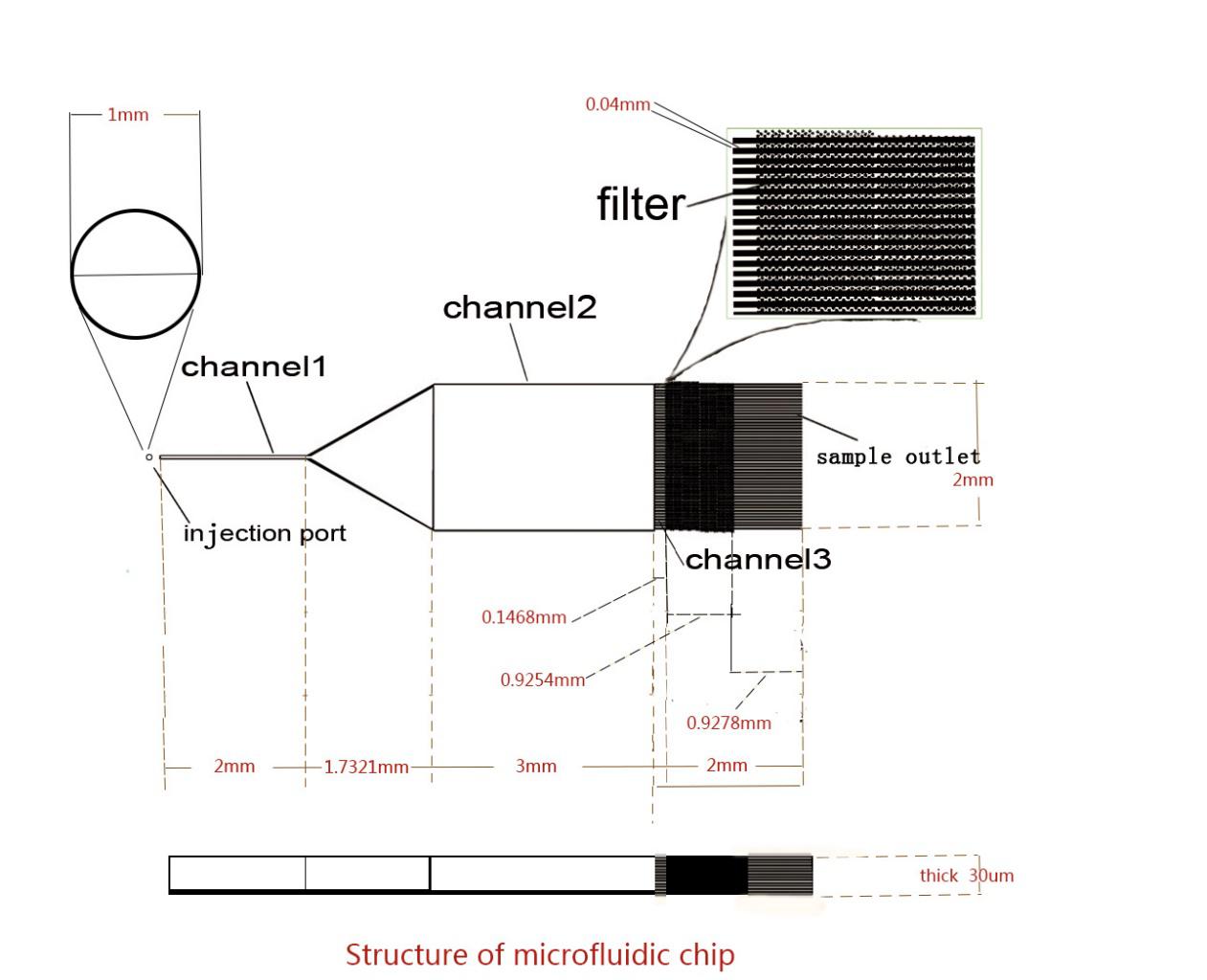
Fig.1 Structure of microfluidic chip.
Just as the figure above, the microfluidic chip is divided into three channels, Channel 1,Channel 2 and Channel 3. The Channel 1 is a narrow channel allows the Bacterial to broth slowly flows through, then the bacteria enterChannel 2, a relatively wide area, also calledthe magnetic deflection area, in whichbacteria will change course under the action of magnetic field we apply. After deflected in the magnetic field, the bacteria will go into Channel 3 divided into fiftysmaller channels with a filter and the bacteria will stay inside under the effect of the filter.After a certain amount of time, we count the bacteria in the small channel under the microscope.Finallywe build a model according the relationship between bacterial number and magnetic field intensity to show the magnetism of bacteria sample.
As for our solution:
Firstly culture 20mL samples about
What’s more in order to make sure our detecting result is accurate, we composed TEM detection to the same sample we used in microfluidic chip. The result of TEM is below, just as we see, the structure of magnetosome is integrated, and there are also many Fe particle around the cell. So we think the sample that we detected contains normal magnetism and mgnetosome structure. Our magnetosome detection is accurate.


Fig2.The magnetosome structure of AMB-1 we cultured. There are distinct magnetosome chains in the cells, and many Fe particles around it.
Fig3.The figure shows the distribution of bacteria sample.
The red broken line shows the distribution of AMB-1 we cultured, the magnetic field interference is on the left of the detection area. And the distribution shows obvious deflection.
Fig4.The figure shows the distribution of bacteria sample.