|
|
| Line 4: |
Line 4: |
| | | | |
| | {| width="100%" | | {| width="100%" |
| - | | colspan="3" | <font color="#333399" size="5" font face="Calibri"> '''Large Phage March - April Notebook: March 15 - March 31 Daily Log'''</font> | + | | colspan="3" | <font color="#333399" size="5" font face="Calibri"> '''Large Phage March - April Notebook: April 1- April 14 Daily Log'''</font> |
| | | | |
| | <br> | | <br> |
| Line 32: |
Line 32: |
| | <font size="4"> '''3/15/13''' </font> | | <font size="4"> '''3/15/13''' </font> |
| | | | |
| - | 3/15/13 - Friday - Bryan Merrill (BDM)
| + | 4/1/13 KS &BDM |
| - | Spreadsheet
| + | Today we did presentations. I talked about DNA packaging in phage. Right now we’re waiting for our phage to come in so we can do some mutagenesis! |
| - | for plasmids, primers, and bacterial strains (and phage) | + | |
| - | Notes
| + | |
| - | on what we found:
| + | |
| - | Largest:
| + | |
| - | Phage G, lytic, capsid, BSL1, generation time = 40 mins.
| + | |
| - | Smallest:
| + | |
| - | RH1 infects Rhodococcus, capsid size of 43 nm, 50kb?
| + | |
| - | Smallest
| + | |
| - | for E. coli: Spreadsheet (Jade) MS2, capsid protein size 137 aa, 27 nm.
| + | |
| - | Well
| + | |
| - | charaterized, Qbeta as well
| + | |
| | | | |
| - | Dissertation
| + | 4/3/13 KS & BDM |
| - | on capsid design – do longer protein sequences correspond to bigger
| + | |
| - | capsids? Are bigger capsids built from
| + | |
| - | bigger monomers or more monomers?
| + | |
| - | T7 – list of all genes, Gp10a and Gp10b (major and minor capsid proteins)
| + | |
| - | Read the thesis/dissertation –
| + | |
| - | 3 Groups: | + | |
| - | Large phage
| + | |
| - | Small phage
| + | |
| - | How to build the capsids (heat shock or self-assembly)
| + | |
| - |
| + | |
| - | Find phage which are well-characterized
| + | |
| - |
| + | |
| - | Find:
| + | |
| - | How
| + | |
| - | phage are used for drug delivery (applications)
| + | |
| - | Find out how capsid sizes change and mutate (targeted mutagenesis)
| + | |
| - | Start by selecting, move towards targeted mutagenesis
| + | |
| - | What are the applications for phage already
| + | |
| - |
| + | |
| - | Cloning, sequence, and expression of the temperature-dependent phage T4 capsid assembly gene 31
| + | |
| - | Modulation of bacteriophage T4 capsid size.
| + | |
| - |
| + | |
| | | | |
| | + | Final Project notes: |
| | + | 1. Watch a science talk and write a paragraph about the presentation. Write a one paragraph critique. |
| | + | T/TH 11:00 and 4:00 |
| | + | 2. Give a presentation on our project so far (15 minutes) |
| | + | 5-10 minutes on the Background |
| | + | -Hypothesis, what is our question, why are we researching this |
| | + | 5-10 minutes methods and results |
| | + | Conclusion- Short, clear bulleted |
| | | | |
| - | Goal:
| + | Things we need: |
| - | Build a library of multiple sizes of phage capsids and
| + | H-Broth |
| - | provide tools to assemble these phage capsids so they can be used for drug
| + | adenine (20ug/ml) |
| - | delivery and nanotechnology
| + | e.coli B bacteria |
| | + | m9+ medium |
| | + | uricil |
| | + | 200ug 5-bromodeoxyuridine, 20ug uridine, 20ug adenine per ml |
| | + | *all work was done in yellow light? |
| | + | chloroform |
| | + | calcium chloride |
| | + | magnesium chloride |
| | | | |
| | + | To do: grow up T1 and T4 |
| | | | |
| - | Look
| + | 4/5/13-KS and BDM |
| - | into:
| + | Today we did a spot test with 11T4, T4Do(s), 12T4r, 40T4r, 40T4 with e.coli BL21. |
| - | Metabolic
| + | Procedure: |
| - | load on bacteria based on capsid size?
| + | We plated .5 mL of an overnight culture of E. coli BL21 in 4.5 mL of LB 1x top agar. When it had hardened, we spot tested 5 uL of the following samples on the plates: 11T4, T4Do(s), 12T4r, 40T4r, 40T4 into 6 sectors of a plate. T4Do came from Dr. Casjens at the University of Utah. We incubated these plates at 37 degrees Celsius. |
| | + | We plated a control of just bacteria. |
| | + | Also, we infected .5 mL of BL21 with 1 uL of the T4Do sample for 20 mins, and then plated it. |
| | + | We started two liquid cultures, each containing 1 mL of BL21. One received 1 uL of T4Do, the other received 10 uL. These were incubated on a shaker at 37C. |
| | | | |
| - | Screening
| |
| - | methods:
| |
| - | Increasingly
| |
| - | harsh environments, less nutrients, smaller phage?
| |
| - | Using
| |
| - | ozone for selection
| |
| | | | |
| - | 3/15/13-Keltzie Smith
| + | 4/8/1/3-BDM and KS |
| - | Today we narrowed in on the phage we are going to use as a starting point for expansion. We found that it wasn’t as easy as we thought to simply look up what would be best for us since there are bits information here and there, but not all in the same location- I suppose this is why we are doing the research that we are- filling a gap! In deciding which to use we took into account genome size as well as how characterized it is. We decided that T4 is the simplest and most effective route to take even though it is technically not the largest phage that infects E. coli.
| + | [[File:Example.jpg|400px]] |
| | | | |
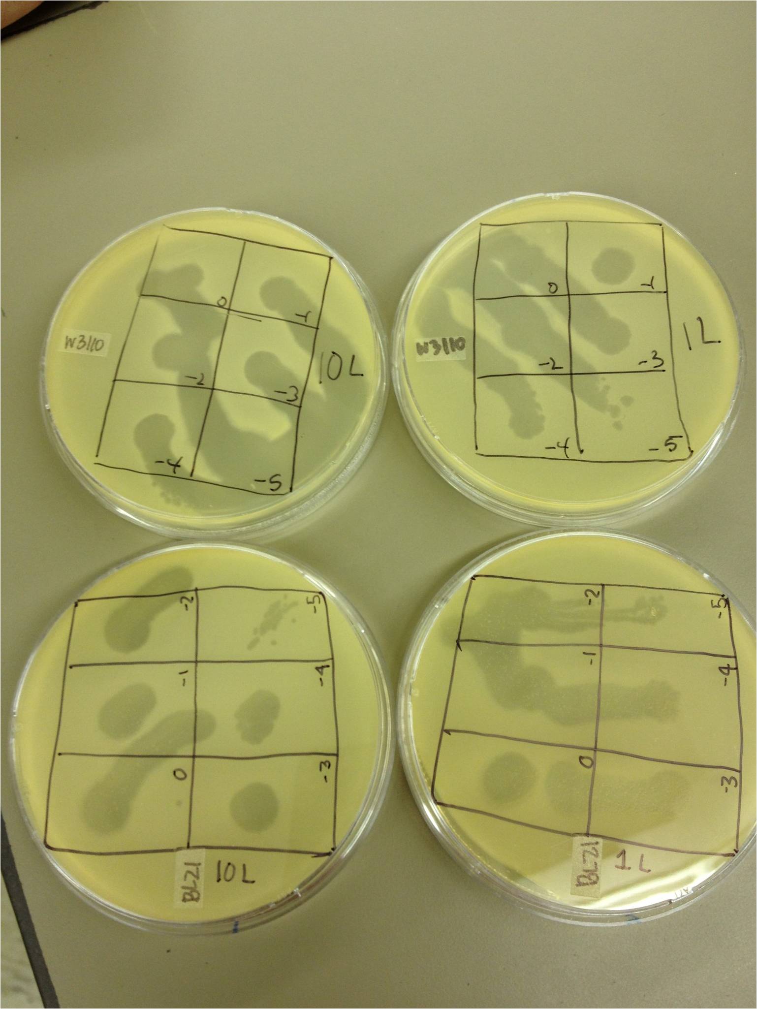
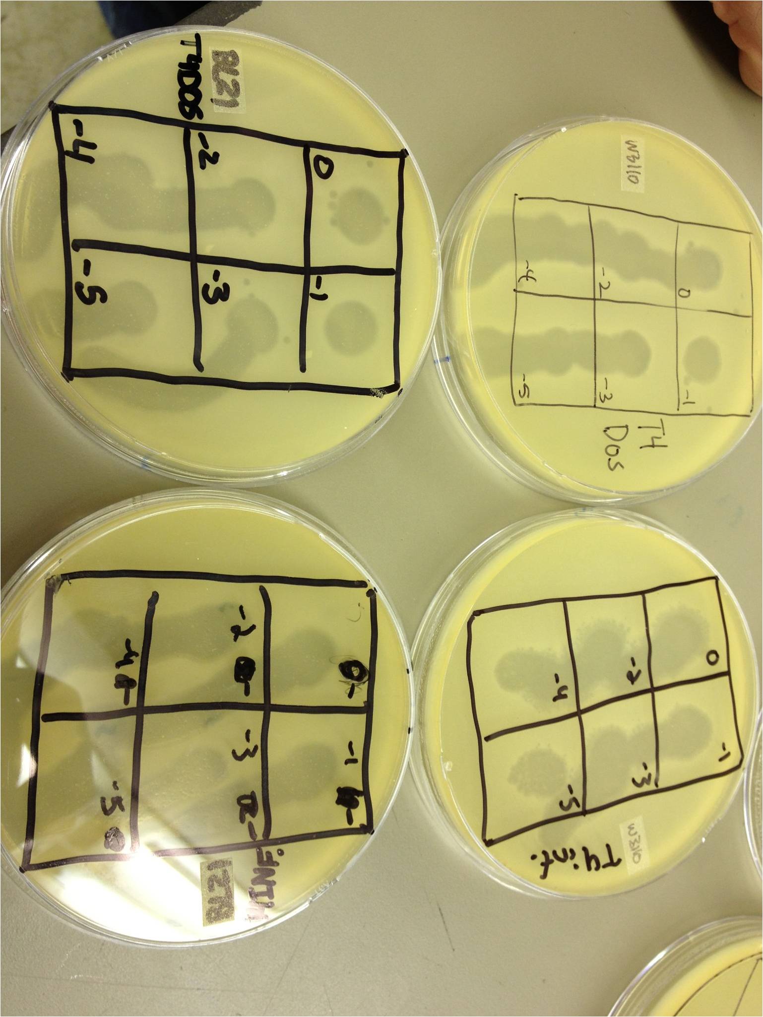
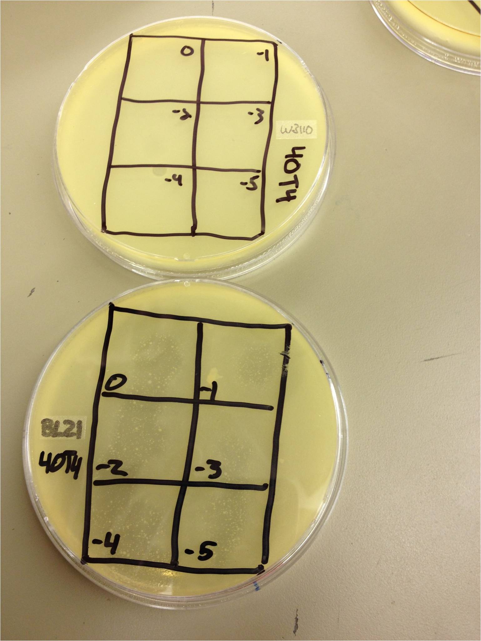
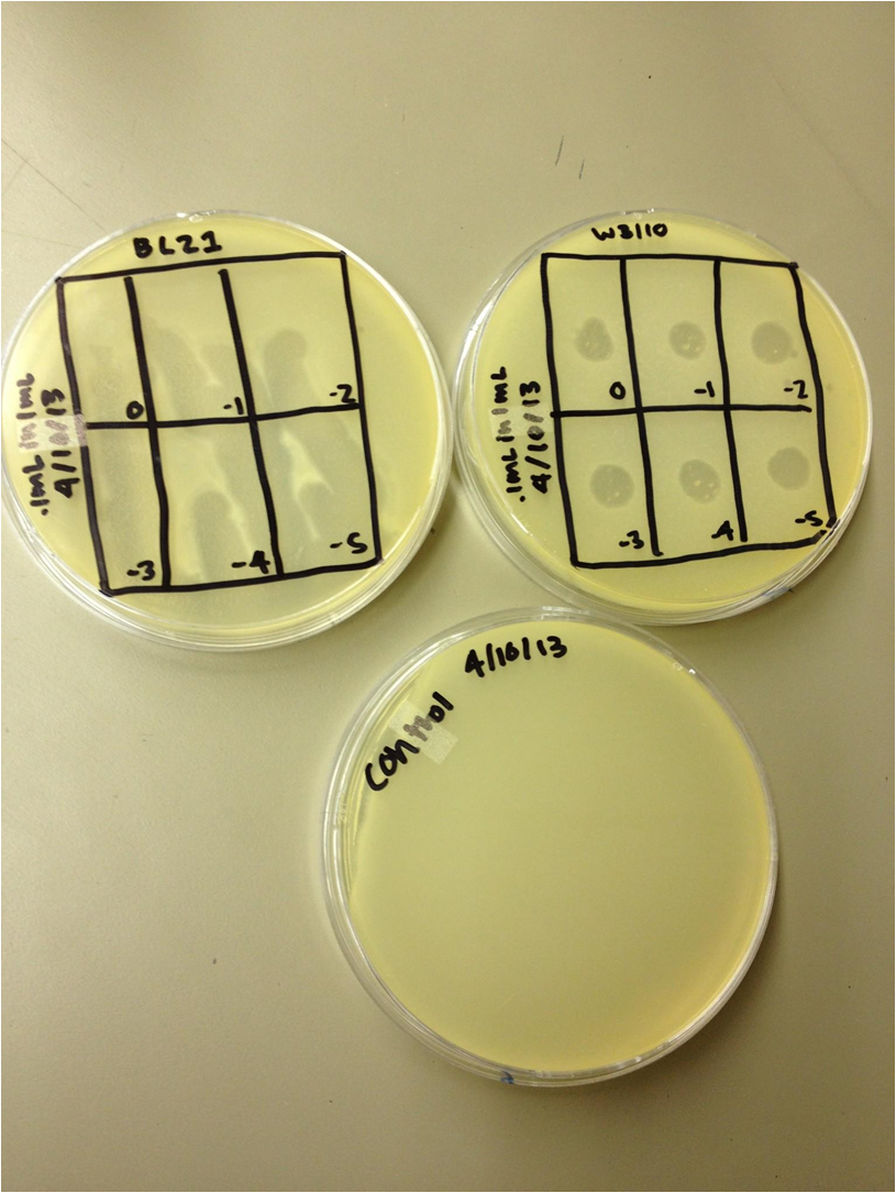
| | + | We performed a phage titer spot test on two bacteria. We used the BL21 and W3110 strains of E. coli. We first put 100 microL of broth into epindorf tubes. We then took 10 microL of 1L, 10L, 40T4, T4DOS, and T4 infected phage and placed it in separate tubes labeled -1. We performed a dilution series taking out 10 microL each time. The last tube we did not remove 10 microL so the total volume was 110 microL. Next we added 4.5 mL of 1x top agar to .5mL of bacteria for both strains and plated it on LB plates. We had previously divided the plates into six sections with the labels 0, -1, -2, -3, -4, and -5. We took the phage from each concentration and spotted it on the plates. We allowed the plates to sit and let the phage soak in. We then placed the plates in the 37 C overnight. |
| | | | |
| - | | + | Results (Wed. 4/10/13) |
| | + | We saw phage plaques in all spots (10^0 through 10^-5) on all bacteria for 10L (10uL of T4Do stock in 1 mL of bacteria grown over the weekend), 1L (1uL same as above), T4Do (stock), and T4 infected plate flood. 40T4 from the fridge grew on BL21 but not W3110. |
| | + | I had started two liquid cultures of 100uL of liquid from the plate flood in 1mL of W3110 and BL21. The W3110 bacteria was visibly lysed (clear); the BL21 bacteria had some snot but was not lysed. |
| | | | |
| | + | Procedure |
| | + | We pelleted the liquid from the two liquid cultures and did a 10^0 through 10^-5 dilution on each, along with a bacterial lawn control. |
| | + | We also started two more liquid cultures of 1 mL from the plate flood in 10 mL of W3110. We will shake these in a 100 mL flask at 37C. |
| | | | |
| | + | [[File:Example2.jpg|400px]] |
| | + | [[File:Example3.jpg|400px]] |
| | + | [[File:Example4.jpg|400px]] |
| | | | |
| | | | |
| | | | |
| - |
| |
| - |
| |
| - |
| |
| - |
| |
| - |
| |
| - |
| |
| - |
| |
| - |
| |
| - |
| |
| - |
| |
| - | Large phage procedures:
| |
| - |
| |
| - |
| |
| - |
| |
| - |
| |
| - |
| |
| - |
| |
| - |
| |
| - | Paper about UV light and backup genes?
| |
| - |
| |
| - |
| |
| - |
| |
| - | Decreasing agar concentration – select for
| |
| - | small plaque sizes
| |
| - |
| |
| - |
| |
| - |
| |
| - |
| |
| - |
| |
| - |
| |
| - |
| |
| - | Find T4 phage and host
| |
| - |
| |
| - |
| |
| - |
| |
| - | Start growing them (archive sample of the
| |
| - | phage)
| |
| - |
| |
| - |
| |
| - |
| |
| - | Determine sequence of the relevant capsid
| |
| - | proteins
| |
| - |
| |
| - |
| |
| - |
| |
| - | Mutate them
| |
| - |
| |
| - |
| |
| - |
| |
| - | Determine the sequence again
| |
| - |
| |
| - |
| |
| - |
| |
| - | Find a way to create just capsids of the larger
| |
| - | size
| |
| - |
| |
| - | <br>
| |
| - |
| |
| - | <font size="4"> '''3/18/13''' </font>
| |
| - | 3/18/13-Keltzie Smith
| |
| - | As of right now our main priority is making sure the T4 we have now is working. Today we started our K12 E.Coli. Next time we’ll see if our T4 is viable, once we see plaques on our plates our next step will be to sequence our phage to determine what we have to start out with an we’ll mutate from there. One thing we are hoping to look deeper into is UV irradiation to select for bigger bacteriophages.
| |
| - |
| |
| - | 18 March 2013 - BDM
| |
| - | 18 March 2013 – Monday – BDM
| |
| - | Notes:
| |
| - | Cholera group
| |
| - | Biofilm – grow a cholera biofilm in the lab to test constructs that have already been created
| |
| - | Plasmid prep – sequencing inserts in plasmid to see if the genes were inserted correctly.
| |
| - | 2 membrane proteins, 2 phosphorylation proteins, 2 genes turned on (RFP and GFP), small RNAs. Need to clone in protein interaction and siRNA degradation.
| |
| - |
| |
| - | Phage group
| |
| - | Protein prep/capsid purification – T7 proteins, requires scaffold protein and capsid proteins to assemble. Other phage, sometimes this doesn’t happen. T4 forms procapsids, but not large ones in vitro. No DNA in capsids – knock out polymerases. Capsid size can depend on environmental factors.
| |
| - | Small phage – Directed and random mutagenesis
| |
| - |
| |
| - | Genetic Control of Capsid Length in Bacteriophage T4
| |
| - | Genetic studies on capsid-length determination in bacteriophage T4
| |
| - | Evolution of Phage Capsid and Genome Size
| |
| - | The Capsid of the T4 Phage Superfamily
| |
| - |
| |
| - | Hendrix, R.W. (2009) Jumbo bacteriophages. Curr. Top. Microbiol. Immunol. 328:229-240
| |
| - |
| |
| - | Lee, K.K., L. Gan, H. Tsuruta, C. Moyer, J.F. Conway, R.L. Duda, R.W. Hendrix, A.C. Steven, and J.E. Johnson (2008) Virus capsid expansion driven by the capture of mobile surface loops. Structure 16:1491-1502
| |
| - |
| |
| - |
| |
| - | <br>
| |
| - |
| |
| - | <font size="4"> '''3/20/13''' </font>
| |
| - |
| |
| - | 3/20/13-Keltzie Smith
| |
| - | Today we learned about phage titering.
| |
| - | pfu's are plaque forming units. We tried to see how competent our phage was, unfortunately we ran into quite a few problems. Much of it due to our lack of experience. We put 10 microliters of phage with 90 microliters of LB and did a 1:10 dilution series. we put 20 microliters of each of the dilution series with .5mLs of E.coli. After we let a twenty minute incubation period pass we added 5 mLs of top agar and plated the phage. With the way everything went, I would imagine there will be a lot of contamination, but I certainly learned quite a bit about the process. Hopefully we see some good growth!
| |
| | � | | � |
| - | 20 March 2013 - BDM
| + | 12 April 2013 - Friday - BDM and KS |
| - | Procedure Description - Titering all stock samples of E. coli phage from Dr. Breakwell’s fridge
| + | The liquid culture titers from last time worked well--there were spots down to 10^-5. |
| - | Purpose - See which phage stocks are viable, and how many PFUs/mL they have.
| + | The liquid culture flasks just looked like normal bacteria--there was no visible lysis. |
| - | Theoretical
| + | |
| - | Protocol - We performed a 1:10 dilution series (10^0 to 10^-5) on each of the stock samples. We infected 0.5 mL of an overnight growth of E. coli K12 with 20 uL of each dilution series tube. After infecting for 20 minutes, we added 5 mL of 1x LB top agar and plated them on LB plates. We let them incubate at 37C overnight.
| + | |
| - | I also tried a spot test of each sample on an E. coli lawn (to check for plaque formation) and on a plain LB plate (to check for phage lysate contamination).
| + | |
| - | Results
| + | |
| - | Actual
| + | |
| - | Protocol - We performed this experiment as we planned on doing it. The agar frequently boiled over in the microwave which made it difficult to use.Next time we will use 2X and mix it right before we need it.
| + | |
| - | Results - None of the samples showed signs of plaques after 24 hours. I will let them incubate one more night. Three of the phage lysates were contaminated.
| + | |
| - | Observations - It is very likely that these phage samples are no longer viable. We ought to make a list of which phages we would like from Dr. Casjens at U of U and start from fresh samples instead of these ones.
| + | |
| - | | + | |
| - | <br>
| + | |
| - | | + | |
| - | <font size="4"> '''3/22/13''' </font>
| + | |
| - | | + | |
| - | 22 March 2013 - Friday - BDM
| + | |
| - | Procedure description - Check plates for any plaques that appeared during the second 24 hours.
| + | |
| - | Identify phage samples we need to request from Dr. Casjens at U of U and send the list to Dr. Grose.
| + | |
| - | We still saw no plaques and a lot of contamination. We’re going to get Dr. Grose a list of the phage we want... probably just T4.
| + | |
| - | | + | |
| - | 3/22/13 Keltzie Smith
| + | |
| - | Today we observed the phage that we plated on Wednesday. There was very little growth of anything so we decided to order some from a professor who has the ones we need. Also Bryan gave us a great tutorial on how to use different programs to compare protein and dna sequences. From here we are going to try to get to know the structure of the phage to better understand how to make point directed mutations.
| + | |
| - | <br>
| + | |
| - | | + | |
| - | <font size="4"> '''3/25/13''' </font>
| + | |
| - | 3/25/13 Keltzie Smith
| + | |
| - | http://www.virologyj.com/content/7/1/356
| + | |
| - | Today we continued research on the t4 capsid. From the article above we discovered that t4 has at least three essential capsid proteins. gp23, gp24 and gp20. With the folding of these essential proteins, chaperone protein systems are involved. For the folding of gp 23 GroEL chaperonin system and phage co-chaperonin gr31 are required. On the capsid of t4 Hoc and Soc “decorate the outside. Hoc(highly antigenic outer capside protein) is a dumbbell shaped monomer at the center of each gp23 hexon, Soc (small outer capsid protein) is a rod shaped moleule that binds between gp 23 hexons. Both Hoc and Soc bind to the capsid after its completion.
| + | |
| - | | + | |
| - | 25 March 2013 - BDM
| + | |
| - | 25 March 2013 – Monday
| + | |
| - | Large Phage
| + | |
| - | Rational design (mutagenesis)
| + | |
| - | Dependent on what we know
| + | |
| - | Genetic selection (UV, agar, etc.)
| + | |
| - | Ask a question, results tell the answer
| + | |
| - | Small phage
| + | |
| - | Vary top agar concentrations to select for smaller phage.
| + | |
| - | Plate T7 on various concentrations of agar; where it fails to form a plaque, start there
| + | |
| - | Purification
| + | |
| - | Found papers for phage purification
| + | |
| - | Liquid culture as well
| + | |
| - | Nanotechnology applications
| + | |
| - |
| + | |
| - | Today: list of reagents
| + | |
| - |
| + | |
| - | Cholera phage and locations:
| + | |
| - | Outbreaks in poorly treated water; Africa, S. America, etc.
| + | |
| - | Most US cases came from travel
| + | |
| - | Find cholera phage
| + | |
| - | | + | |
| - | Biofilms
| + | |
| - | Proteosomes – degrade proteins
| + | |
| - | Amylases – degrade polysaccharides
| + | |
| - | All biofilms are different
| + | |
| - | Sample colonies and determine ratios in the biofilm
| + | |
| - | Plant extracts worked
| + | |
| - | Start growing cholera, see if biofilm comes (incubate at various temperatures)
| + | |
| - |
| + | |
| - | Recipes:
| + | |
| - | LB 2x top agar
| + | |
| - | 10g LB broth
| + | |
| - | 4g agar
| + | |
| - |
| + | |
| - | 10 of 16 genes for prohead formation are essential.
| + | |
| - |
| + | |
| - | GroEL (host-encoded)
| + | |
| - | T4 gp31 can substitute for GroES which binds to GroEL
| + | |
| - |
| + | |
| - | GroEL-gp31 helps gp23 fold (major capsid protein)
| + | |
| - |
| + | |
| - | gp22 and gp21 is part of the scaffold, as well as gp23 - assembled onto initiator (portal vertex) which is 12-mer of gp20 in a ring.
| + | |
| - |
| + | |
| - | Pentamers of gp24 are placed at other 11 vertices.
| + | |
| - |
| + | |
| - | T4 protease (gp21) degrades scaffold and cleaves head proteins (23 and 24) creating space.)
| + | |
| - |
| + | |
| - | DNA packaging forms the initiated small particle
| + | |
| - | | + | |
| - | | + | |
| - | <br>
| + | |
| - | | + | |
| - | <font size="4"> '''3/27/13''' </font>
| + | |
| - | | + | |
| - | 3/27/13 KS
| + | |
| - | I am going to be going deeper into studying about t4 capsid assembly including all the proteins that are involved in it. Hopefully this will give a better look into how we can possibly change things to make a bigger capsid.
| + | |
| - | | + | |
| - | 27 March 2013 - BDM
| + | |
| - | 27 March 2013 – Wednesday
| + | |
| - | Gp40 embeds in membrane
| + | |
| - | Gp20 attaches to Gp40
| + | |
| - | Tons of gpalt, gp21, gp67, gp68, IPI, IPII, IPIII attach
| + | |
| - | Gp22 surrounds it
| + | |
| - | Gp23 and 24 coat the inside
| + | |
| - | Gp21 is activated, everything breaks down and disperses
| + | |
| - |
| + | |
| - | Giant capsids are produced under heavy mutagenic pressure, UV irradiation, and always have multiple genomes in them. Mutagens typically have mutations in gp23.
| + | |
| - |
| + | |
| - | To do:
| + | |
| - | Compile list of reagents needed to mutagenize large phage
| + | |
| - | Put together presentation about the process of doing this
| + | |
| - | | + | |
| - | | + | |
| - | <br>
| + | |
| - | | + | |
| - | <font size="4"> '''3/29/13''' </font>
| + | |
| - | | + | |
| - | 3/29/13 KS
| + | |
| - | Today we spent class researching for our presentations for Monday. I have been working on the mechanism that t4 uses to package the DNA to see how we can take extra DNA and have the t4 encapsulate it to create a larger phage that we want. This is also an important thing to know concerning how they package medicines in there.
| + | |
| | | | |
| - | Friday 29 March 2013- BDM
| + | Today we pelleted and filtered the liquid cultures and worked on our presentations for Monday. |
| - | I prepared my part of the presentation for Monday before class today. I missed most of class but was excused by Dr. Grose.
| + | [[File:Example5.jpg|400px]] |
| | | | |
| | <br> | | <br> |
 "
"
