Team:BYU Provo/Notebook/SmallPhage/Winterexp/Period2/Exp/4.5 T7 phage viability assay
From 2013.igem.org
| Small Phage March - April Notebook: Experiments
| ||
|
|
4.5 T7 Phage Viability Test
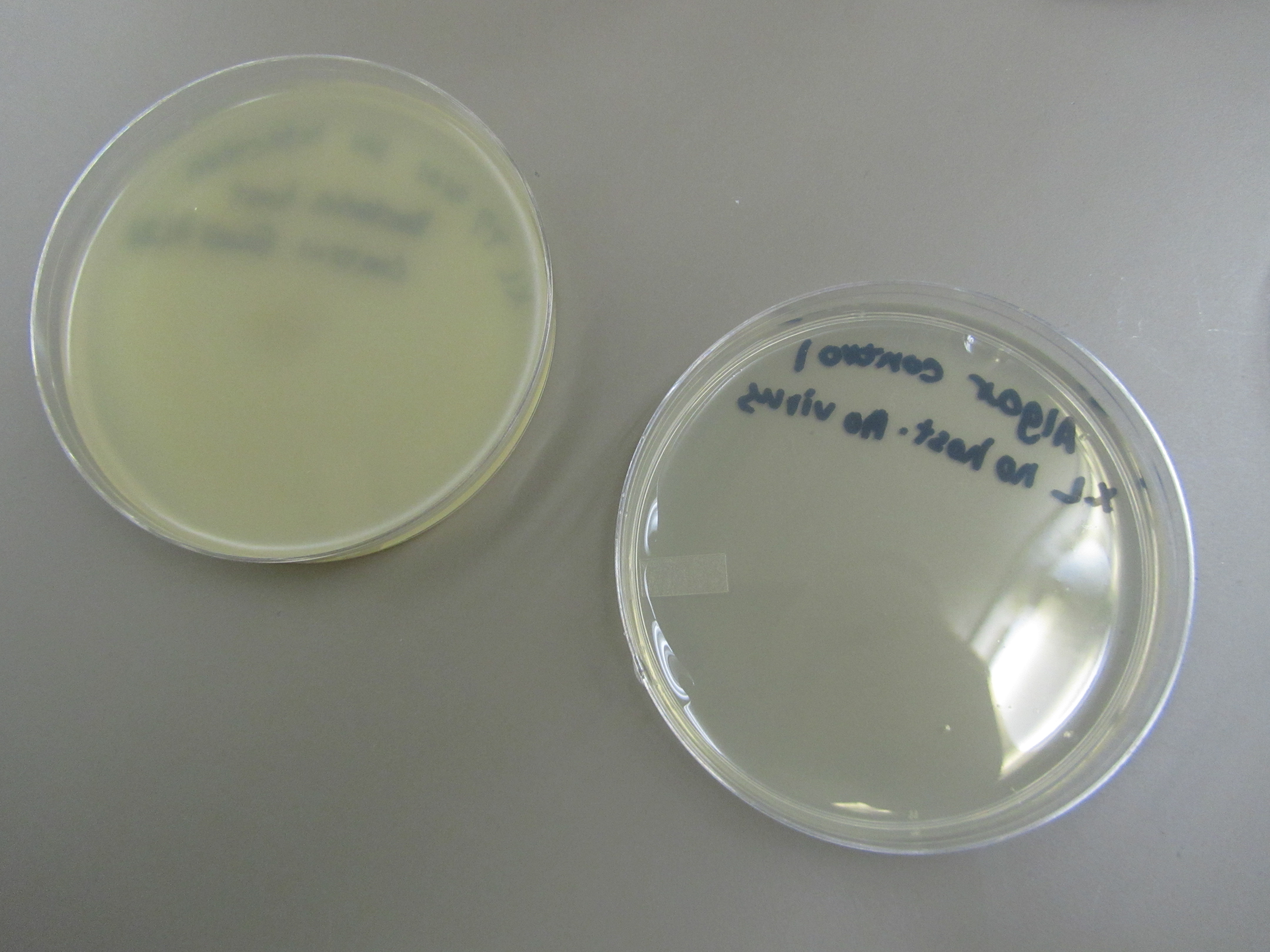
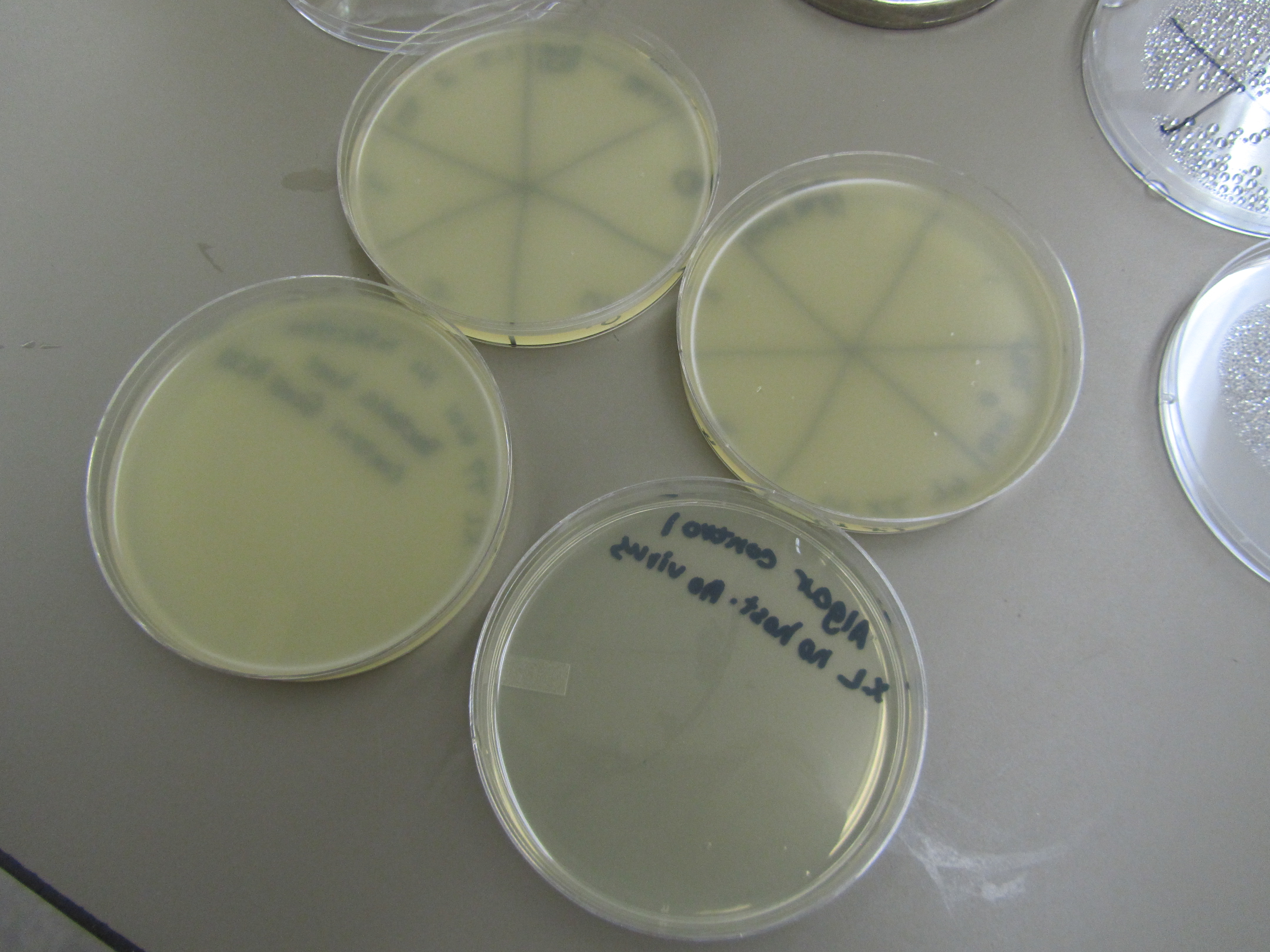
I. Purpose - To determine if our phage are viable - Practice phage titering and other experimental techniques II. Expercted Results - We hope to see plaques in a lawn of E. coli (confluent growth). This would indicate where phage replicated, lysed bacteria, and are viable. III. Protocol - We filled 5 test tubes with 90 microliters of LB each. - We added 10 microliters of T7 4104 to the first test tube and then mixed. - We created a dilution series by taking 10 microliters of the first tubes mixed solution and added it to tube 2, and followed the same procedure for each test tube down the line to tube 5. - We combined 10mL LB, 10mL top agar, and 2mL E. Coli BL21 in a 50mL plastic centrifuge tube. We plated 5mL of this onto 3 plates. - Wait for top agar (containing E. coli BL21) to solidify before proceeding to spot test. - Divide each plate into 6 sections, labeled 0-5. Spot 5μL of liquid from each vial unto different sections of the plate. Vial and section should correspond in number. - Two plates were used as control - Agar control: 1:1 top agar/LB -> 5mL plated. This is to make sure that top agar and LB are not contaminated - E coli control: 5mL of the combined solution from the centrifuge tube (2-i step) were plated. This is to make sure the bacteria stock was not contaminated - Check up on phage + bacteria viability in 24 hours. IV) Results (April 6 at 4: 45pm) 1) The two control plates (Left -> E. Coli control; Right ->Agar control) No contamination observed on these two plates. This confirms that there is no contamination in Agar, LB, or E. Coli stock. Uniform lawn of E coil BL21 observed in E coli control plate. This suggests that our E coli is growing normally 2) Spot test + control plates (Top -> 2 spot test; bottom left -> E coli control; bottom right -> Agar control) No phage clearage is observed. Some small dots on agar can be seen, but they are too small to be resulted from phage lysing E coli. It’s more likely that they are the result of either residue air bubbles or puncture marks made by pipet tips during the spot test. V) Conclusions - E coli BL21 is viable, but the T7 4104 phage does not form plaques on BL21. Or we did not use a big enough volume of phage stock solution to observe a significant lysate. - Another reason for why we did not see phage plaque is that we used the mutant phage without the complete genome: T7 4110 vs T7* -> T7* might actually be the WT phage. VI) Proposed next step 1) Perform the same experiment with a larger volume of phage stock solution. 2) Test T7 phage survival on E coli W3110.
| |
 "
"

