|
- Small Phage
- March-April
- May-June
- July-August
- September-October
|
5.20 T7 Minor Capsid Protein PCR
I) Purpose
- To isolate the T7 minor capsid protein
II) Expected Outcome
- We expect to amplify the T7 minor capsid protein. This can be visualized by gel electrophoresis: there should be one band that matches the number of base pairs in the minor capsid protein.
III) Reagents Used
- ddH2O; Thermo Taq buffer; dNTPs; primer; BI257; BI258; Taq polymerase
IV) Procedure
1) Isolating DNA (5.20)
- - Put 5 ul of phage stock and 45 ul of ddH20 in one PCR tube and 10 ul of phage stock and 40 ul of ddH20 in another PCR tube.
- - Boil for 12 minutes in the PCR machine.
- - Remove the tubes from the PCR machine and shake the tube. Centrifuge it for 1 minutes at top speed.
- - Keep on ice. DNA should be in the supernatant.
2) PCR (5.20)
- - To a centrifuge tube, add
- 120 ul ddH20
- 15 ul 10X TAQ Buffer
- 4.5 ul 10mM dNTP's
- 3 ul of forward primer
- 3 ul of reverse primer
- - Mix well
- - Label 3 PCR tubes "C" (Control), "5" (5 ul of phage stock), and "10" (10 ul of phage stock).
- - Add 2 ul of template DNA from the supernatant of step 1 into their respective tubes (nothing in the control tube).
- - Add 1.5 ul of TAQ Polymerase into the centrifuge tube (master mix).
- - Pipette 48 ul of master mix into each PCR tube.
- - Run 35 cycles with temperatures of 95 C, 50 C, and 72 C with an extension time of 1.5 minutes.
- - Leave overnight at 4 C.
- - Remove and place in the freezer (5.21 at 9:45am).
3) Check with Agarose Electrophoresis (5.22)
- - Make a standard 1% gel using 75 ml of 1X TAE buffer and 0.75 grams of agarose (regular agarose, not low melt). Put into the microwave for about 90 seconds or until the agarose is completely dissolved. Pulsing the microwave may be necessary to prevent boiling over
- - Let the flask cool until it isn't burning hot. Add 1 drop of ethidium bromide and swirl to mix.
- - Pour the liquid onto the gel bed and let it cool. Insert the appropriate sample comb (need 4 slots).
- - Move the gel into the proper orientation (DNA runs towards the red electrode) in the gel box, cover the gel with 1X TAE buffer and load 8 ul of each of the PCR products with 3 ul of loading dye mixed in. Add a DNA ladder as a reference. Turn on the power supply and run at 150-175 volts. Let it run for 25 minutes.
- - Visualize the gel in an Alphaimager.
4) Sequencing (5.29)
- - Two PCR tubes were prepared
- iGem T7 F: 2uL of "5" PCR product + 1uL of forward primer (BI257)
- iGem T7 R: 2uL of "5" PCR product + 1uL of reverse primer (BI258)
V) Results
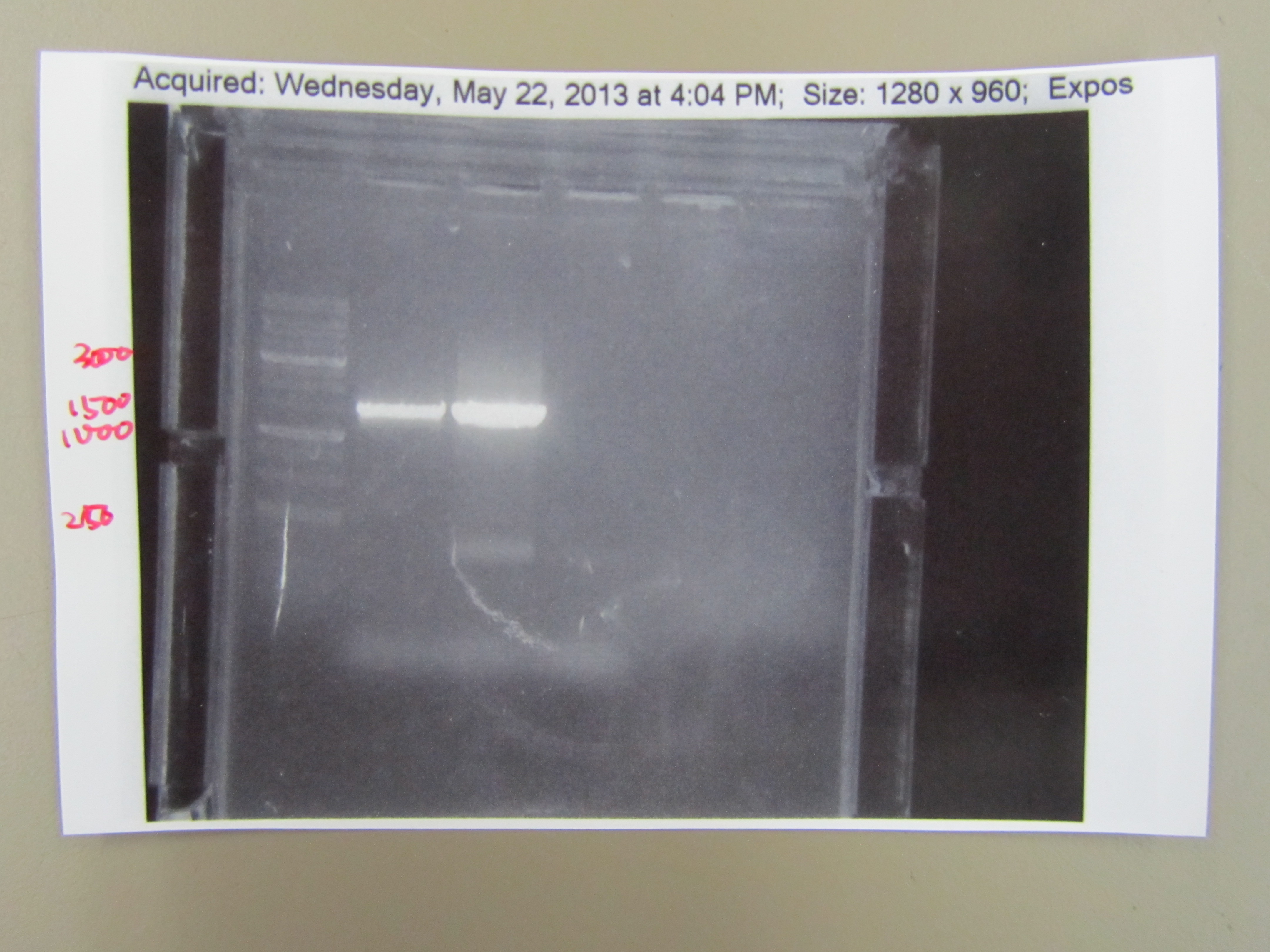
- The bands from the "5" and "10" PCR tubes showed bands at around 1600 base pairs. This confirms that proper PCR was accomplished and there was no contamination. Also, the "10" band was brighter than the "5" band and the control slot had no bands.
- The sequencing results didn't have very high fidelity, but still showed that it was the T7 capsid protein.
|
 "
"
