|
- Small Phage
- March-April
- May-June
- July-August
- September-October
|
7.29 Mutagen Concentration Test - Sixth Protocol
I) Purpose
- To test the sixth protocol for applying 5-bromodeoxyuridine and inducing mutation. We also hope to see if using LB or M9 is better for the mutagen process.
II) Expected Outcome
- A decrease in phage viability with increasing mutagen concentration.
- A few random mutations that produce smaller and larger phage that can be isolated.
- M9 media should allow E. Coli BL21 to take in more of the mutagen so there should be more mutations in T7 phage, resulting in a smaller titer when compared with LB media.
III) Reagents Used
- 5-bromodeoxyuridine stock in freezer labeled T7 (10mg/mL)
- 50mL of M9+ prepared by the large phage group
- Uracil solution (2.5mg/mL)
- Adenine solution (5mg/mL)
IV) Procedure
1) Overnight (The day before) (7.28)
- Add 5mL of LB into a test tube and add 1 colony of E. Coli BL21 into the tube using a wooden stick.
2) Applying the mutagen (7.29)
- Label 10 test tubes LB:C, LB:0, LB:100, LB:200, LB: 500, M9:C, M9:0, M9:100, M9: 200, and M9: 500. Add 9.8mL of LB, 40ul of adenine solution into each test tube. Then add 200ul of BL21 overnight into each test tube. Incubate on the shaker at 37 Celsius.
- Make the M9+ suspension medium and supplement it with the appropriate amount of uracil and adenine. In this case, 200uL of adenine solution and 400uL of uracil solution was add 50mL of M9+.
- Make a LB/adenine/uracil solution by adding 200uL of adenine solution and 400uL of uracil solution to 50mL of LB.
- Remove all the test tubes off the shaker after 2 hours. Centrifuge all the tubes at 4000rpm for 10 minutes at 7 Celsius. Discard the supernatant using pipettes. For the LB tubes, add 10mL of the LB/adenine/uracil solution into each tube with a pipette, being sure to pipette up and down to resuspend the bacteria. For the M9 tubes, add 10mL of the M9 solution into each M9 labeled tube with a pipette and resuspend the bacteria.
- To each tube, add the corresponding amount in ul of 5-bromodeoxyuridine, a mutagen, to each test tube with a 100, 200, or 500. (Ex: Add 100ul of mutagen to LB:100 and M9:100.)
- Take 1mL from the LB:C tube and pipette it into a cuvette. Using 1mL of LB in a cuvette, blank the spectrophotometer at 600 OD. Then measure the absorbance of the LB:C cuvette.
- Place all the tubes on the shaker at 37 Celsius for 30 minutes.
- Remove the tubes and add 10ul of T7 phage from the "10ul 7.19 stock" to each tube, except for the tubes with a "C" on them. (There should be a 1:10 phage to bacteria concentration.)
- Incubate all the tubes on the shaker at 37 Celsius for 80 minutes.
- Remove all the tubes (place tubes with a "C" to the side; they are no longer needed) and add 1mL of chloroform to each. Gently shake each tube and centrifuge it at 4000rpm for 10 minutes at 7 Celsius. Remove the supernatant from each tube with a pipette and place it in a new tube with the same label. Be careful not to get the chloroform or bacteria when you remove the supernatant.
- Store the supernatants at 4 Celsius.
3) Spot test to determine phage concentration (7.30-8.1)
- Dilution series between -2 and -6 was performed for each of the 8 samples (M9 and LB each at 0ug, 100ug, 200ug, and 500ug).
- Spot test was performed by plating 0.5mL of E coli BL21 liquid culture overnight and 5mL of x1 top agar, and spotting 5uL of each dilution sample.
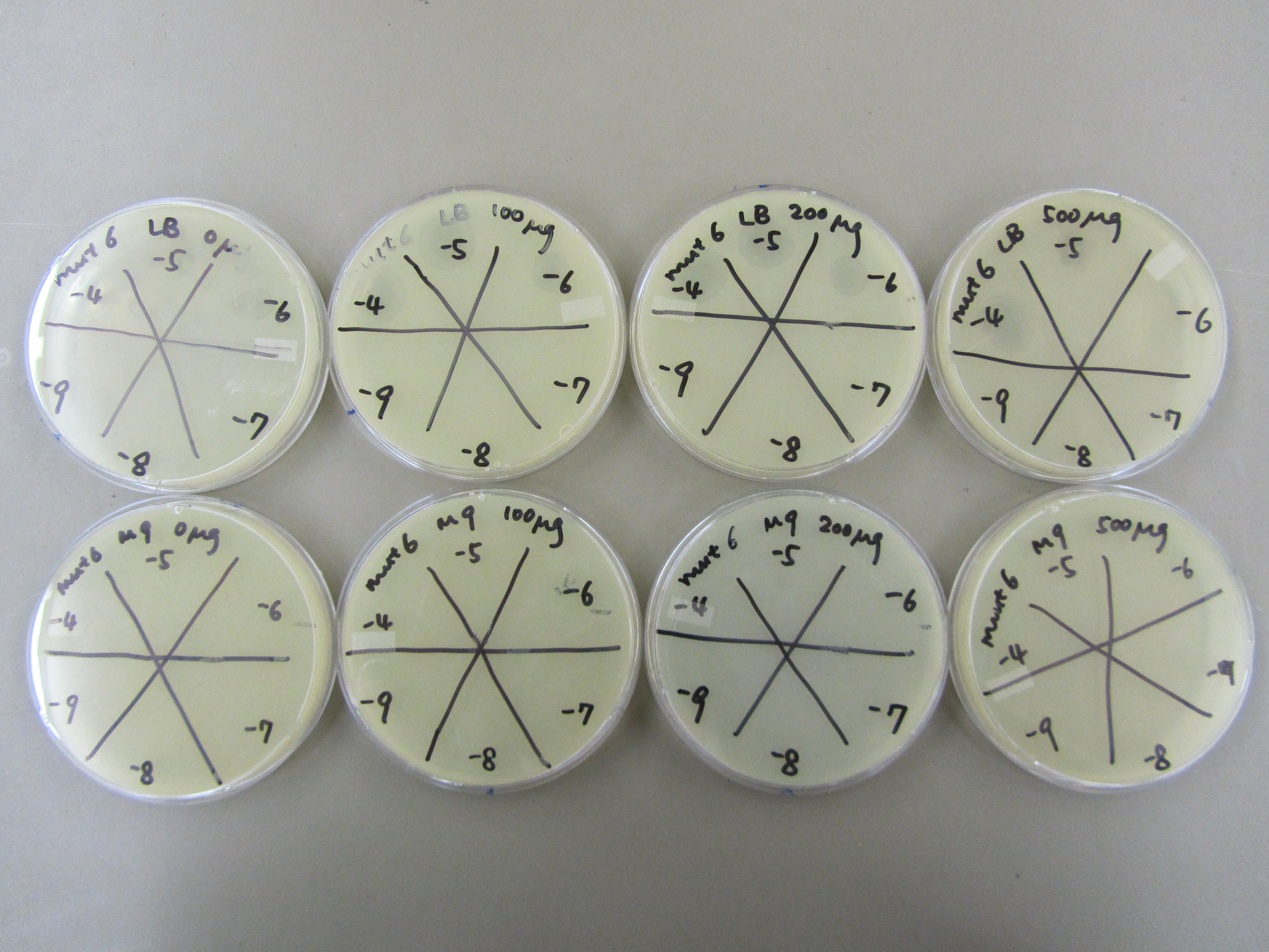
4) Spot test to determine phage concentration (8.2 and 8.5)
- Each sample was further diluted to -7, -8, and -9.
- Spot test was performed by plating 0.5mL of E coli BL21 liquid culture overnight and 5mL of x1 top agar, and spotting 5uL of each dilution sample (-4 through -9).
5) CsCl gradient
- The mutagenized phage was handed over to the phage purification team for CsCl gradient separation. Due to multiple factors (gradient design not optimal, switch in CsCl vendor, etc). All phage banded at the top of the gradient. Adjustments to gradient design will be made based on the results from this experiment.
V) Results
2) Applying the mutagen
- The OD reading was 0.275. After another 30 minutes of incubating, the bacterial concentration should be about 2E8. This means we wanted to add 2E7 phage, which is roughly 10ul of the "10ul 7.19" stock.
3) Spot test to determine phage concentration
- Every plate had contamination. The contaminant colonies are randomly spread out on the plates, implying that most likely the top agar used was contaminated. We also observed that these colonies are bigger than usual at places where phage plaques should be observed. This can be explained by the fact that T7 will have lysed BL21 at places where the plaques were forming, leaving the spot open for contaminant growth.
- Even with contamination, we can tell that most of the plates formed plaques at -6. To verify this result we will further dilute the samples and perform another spot test.
4) Spot test to determine phage concentration
- Experiments in LB produced a much higher titer of phage as compared to M9. This could be due to phage death during mutagenesis or its storage in the fridge. But for the CsCl gradient to work we need a higher titer of phage, at least 108 pfu/mL. Thus we'll use LB from now on.
VI) Conclusion
- The purpose of this experiment was the determine whether M9 of LB will better support the mutagenesis of phage. Although we predicted that the M9 would be a better choice, considering phage concentration, we will have to use LB instead.
- The second purpose of this experiment was to generate mutagenized phage for the phage purification team. Their experiments this time did not go so well, thus we will proceed with 8.14 Mutagen Concentration Test - Seventh Protocol to produce more phage for them to work with.
|
 "
"