Team:ITB Indonesia/Project/Aflatoxin
From 2013.igem.org

Aflatoxin Whole Cell Biocensor
Aflatoxin Whole Cell Biocensor is a system to detect the presence of aflatoxin in the environment by using E. coli strain BL21 as a chassis. There are two modules are used to compile the aflatoxin sensor systems. Module I is a module to activate aflatoxin. Module II is a module which is used as a reporter system utilizing the SOS response in E. coli cells. When E. coli cells exposed to aflatoxin, aflatoxin will enter the cell and activated by the system in module 1 form the active molecule. This molecules are not stable and will attack the DNA. DNA damage will activates the system at the module II to produced green fluoresence protein as an output from module II system.
Modul I. Activation Aflatoxin B1 (AFB1) become Exo-AFB1-8,9-epoxide
Alfatoxin is a mycotoxin that is carcinogenic and mutagenic. To be able to cause mutations or DNA damage, AFB1 must undergo activation by Cytocrhome P450 prior to Exo-AFB1-8,9-epoxide. Exo-AFB1-8,9-epoxide is the active form of AFB1 which can cause damage to DNA.
Cytochrome P450 is a superfamily proteins that catalyze the activation of a variety of organic compounds such as intermediate compound in the metabolism proses, or xenobiotic compounds. Cytochrome P450 found in almost all domains of life except in E.coli. Cytochrome P450 3A4 (CYP3A4) is one member of the cytochrome P450 superfamily of proteins derived from human. CYP3A4 has a native function to activate xenobiotic compounds in the human body, including aflatoxin [1].
In this module synthetic CYP3A4 gen [BBa_K1064004] from humans was used. CYP3A4 has a high efficiency to convert AFB1 into its active form (Exo-AFB1-8,9-epoxide). Doesn't like other types such as CYP1A5 that much change AFB1 into Endo- AFB1-8,9-epoxide (not active) or even converted to AFM1 [2]. Naturally CYP3A4 is a membrane protein , so we cut the signal peptide to prevent CYP3A4 protein translocated to the cell membrane after translation. Localization CYP3A4 in cytosol is intended to only activate AFB1 that had enter the cell so it'll reducing the error in measurement aflatoxin concentration in the environment. The activation of AFB 1 into Exo-AFB1-8,9-epoxide has been modeled.

Picture 1. Ilustration of Modul I
Modul 2. Utilisizing SOS Response to Detection the Presence of Aflatoxin
Aflatoxin activated into Exo-AFB1-8,9-epoxide by CYP3A4 as a catalyst. Exo-AFB1-8,9-epoxide is an unstable compound and could not be isolated. The compound can adduct DNA at guanine bases produce a compound trans-8,9-dihydro-8-(N7-guanyl)-9-hydroxyaflatoxin B1 and intercalated in DNA [3]. On the E. coli, DNA adduction by Exo-AFB1-8,9-epoxide cause transversion mutation of nucleotide bases G à T cell triggering of the SOS response.

Picture 2. DNA adduction by Exo-AFB1-8,9-epoxide
SOS Response is a response of E. coli to DNA damage. Mechanism of SOS respone to repair damaged DNA involving multiple gene. There are two types of proteins that are involved in the mechanism of the SOS response in E. coli, the LexA which is a repressor protein and RecA which for reducing the level of expression of the gene encoding LexA
Under normal condition, the LexA Protein which is a dimer protein binds to the operator and represses the transcription process of genes that play a role in DNA repair. When cells undergo UV exposure or mutagenic compounds that cause DNA damage, DNA damage will be detected when the process of DNA replication occurs. DNA pol III will fail at replicating DNA, produce a ssDNA. At that time, RecA protein binds to another protein with ssDNA and continuing DNA replication via homologous recombination. When RecA protein binding to ssDNA the protein becomes active to cleavage protein LexA on operator lead to transcription genes that play a role in the repair of damaged DNA [4]
SOS response system on module II is utilized to regulate the expression of the gene encoding GFP. DNA Damage due to attack by Exo-AFB1-8,9-epoxide would trigger the SOS response, for that it we utilizing the SOS promoter that regulates expression of the gene encoding GFP, so the presence of aflatoxin can be detected.

Picture 3. Ilustration of Modul 2.

Picture 4. How System Works
Results of agarose gel electrophoresis of restriction analysis and PCR results showed that the construct of the system on module 2 has succeeded

Picture 5. Electroforegram from Analisis restriction construct pSOS+GFP
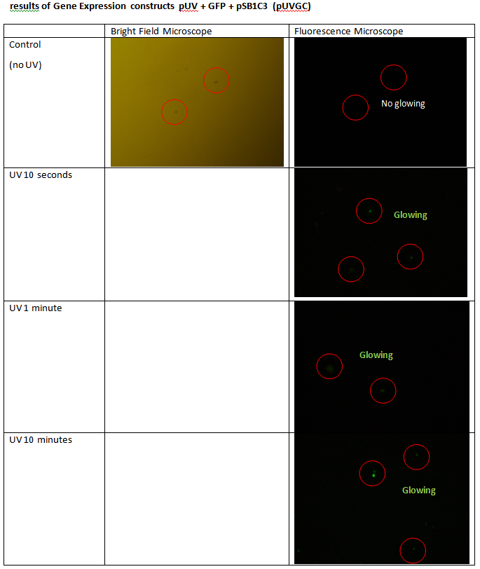
To check that the system in our construct running or not, we test the construct by exposured E. coli containing plasmid constructs in UV light. Results of fluorescence microscopy observations showed that the cells were carried out for exposure to UV light produces a green fluoresence whereas control cells that are not exposed to UV light do not produce fluoresence. It shows that the system in modul 2 runs with a good to take the DNA damage signal as a signal for GFP gene expression
results of Gene Expression constructs pSOS + GFP + pSB1C3 (pSOSGC)
results of Gene Expression constructs pUV + GFP + pSB1C3 (pUVGC)
[1]. Guengerich FP (January 2008). "Cytochrome p450 and chemical toxicology".Chem. Res. Toxicol. 21 (1): 70–83
[2]. Rawal, Summit., S. M. Yip, Shirley., Roger A. Coulombe, Jr.2010. Cloning, Expression and Functional Characterization of Cytochrome P450 3A37 from Turkey Liver with High Aflatoxin B1 Epoxidation Activity. Chem. Res. Toxicol. 2010, 23, 1322–1329
[3]. Michael P. Stone, Surajit Banerjee, Kyle L. Brown, and Martin Egli. Chemistry and Biology of Aflatoxin-DNA Adducts
[4]. Shimoni Y, Altuvia S, Margalit H, Biham O (2009) Stochastic Analysis of the SOS Response in Escherichia coli. PLoS ONE 4(5): e5363. doi:10.1371/journal.pone.0005363
 "
"

