Team:Tokyo Tech/Experiment/Crosstalk Confirmation Assay
From 2013.igem.org
| Line 61: | Line 61: | ||
<h3>4-3. Protocol </h3> | <h3>4-3. Protocol </h3> | ||
<h2> | <h2> | ||
| - | < | + | <blockquote> |
| - | 1. O/N -> FC -> Induction< | + | <b>1. O/N -> FC -> Induction</b> |
| + | </blockquote> | ||
| + | |||
<p><blockquote> | <p><blockquote> | ||
1.1 Prepare overnight culture of each cell (GFP posi, GFP nega, sample) at 37°C for 12 h. <br> (=> O/N) | 1.1 Prepare overnight culture of each cell (GFP posi, GFP nega, sample) at 37°C for 12 h. <br> (=> O/N) | ||
| Line 73: | Line 75: | ||
</blockquote></p> | </blockquote></p> | ||
<p><blockquote> | <p><blockquote> | ||
| - | 1.4 Dilute the flesh culture in 1:5 by the following conditions: | + | 1.4 Dilute the flesh culture in 1:5 by the following conditions: |
| - | ...LB (3 mL) + antibiotics (Amp 50 microg/mL + Kan 30 microg/mL) + 5 microM 3OC6HSL (3 microL)< | + | <blockquote>...LB (3 mL) + antibiotics (Amp 50 microg/mL + Kan 30 microg/mL)<br> + 5 microM 3OC6HSL (3 microL)</blockquote> |
| - | ...LB (3 mL) + antibiotics (Amp 50 microg/mL + Kan 30 microg/mL) + 5 microM 3OC12HSL (3 microL)< | + | <blockquote>...LB (3 mL) + antibiotics (Amp 50 microg/mL + Kan 30 microg/mL)<br> + 5 microM 3OC12HSL (3 microL)</blockquote> |
| - | ...LB (3 mL) + antibiotics (Amp 50 microg/mL + Kan 30 microg/mL) + 5 microM DMSO (3 microL) | + | <blockquote>...LB (3 mL) + antibiotics (Amp 50 microg/mL + Kan 30 microg/mL)<br> + 5 microM DMSO (3 microL)</blockquote> |
| - | </blockquote> | + | |
</blockquote></p> | </blockquote></p> | ||
| + | |||
<p><blockquote> | <p><blockquote> | ||
1.5 Incubate the flesh culture of diluted inducer cell for 4 h at 37°C.<br> (=> Induction) | 1.5 Incubate the flesh culture of diluted inducer cell for 4 h at 37°C.<br> (=> Induction) | ||
</blockquote></p> | </blockquote></p> | ||
<br> | <br> | ||
| - | + | ||
| - | + | ||
<blockquote> | <blockquote> | ||
| - | + | <b>2. Measurement (Flow cytometer)</b> | |
| - | 2. | + | |
| - | + | ||
| - | + | ||
| - | + | ||
| - | + | ||
| - | + | ||
</blockquote> | </blockquote> | ||
| - | </p> | + | |
| + | <p><blockquote> | ||
| + | 2.1 Measure all samples' OD600. | ||
| + | </blockquote></p> | ||
| + | <p><blockquote> | ||
| + | 2.2 Dilute all samples with 1X PBS to keep OD600 in the range from 0.2 to 0.5. | ||
| + | </blockquote></p> | ||
| + | <p><blockquote> | ||
| + | 2.3 Take 1 mL (from all samples) into a disposal tube (for flow cytometer). | ||
| + | </blockquote></p> | ||
| + | <p><blockquote> | ||
| + | 2.4 Centrifuge them at 9000 g, 4°C, 1 min. and take their supernatant away. | ||
| + | </blockquote></p> | ||
| + | <p><blockquote> | ||
| + | 2.5 Suspend all samples with 1 mL 1X PBS. | ||
| + | </blockquote></p> | ||
| + | <p><blockquote> | ||
| + | 2.6 Measure all samples. | ||
| + | </blockquote></p> | ||
| + | <p><blockquote> | ||
| + | 2.7 Save and organize data. | ||
| + | </blockquote></p> | ||
</h2> | </h2> | ||
<h1>5. Result of the assay </h1> | <h1>5. Result of the assay </h1> | ||
Revision as of 14:13, 27 September 2013
Crosstalk Confirmation Assay
Contents |
1. Introduction
Our goal in this project is to construct a system to suppress crosstalk by 3OC12HSL-LasR complex on lux promoter. We thought that we could prove that our system precisely works only after we obtain data of crosstalk happening by ourselves. Therefore, we confirmed that crosstalk is really happening by the following assay.
2. Summary of the experiment
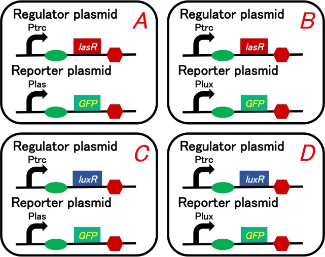
Our purpose is to confirm 3OC12HSL-LasR complex really activates lux promoter. We prepared four plasmids shown in below (Fig. 3-1-1). We checked what would happen when we add intercellular molecules 3OC6HSL and 3OC12HSL.
We prepared twelve conditions as follow.
A-1) Culture containing Ptrc-lasR and Plas-GFP cell with 3OC6HSL induction
A-2) Culture containing Ptrc-lasR and Plas-GFP cell with 3OC12HSL induction
A-3) Culture containing Ptrc-lasR and Plas-GFP cell with DMSO ( no induction)
B-1) Culture containing Ptrc-lasR and Plux-GFP cell with 3OC6HSL induction
B-2) Culture containing Ptrc-lasR and Plux-GFP cell with 3OC12HSL induction
B-3) Culture containing Ptrc-lasR and Plux-GFP cell with DMSO (no induction)
C-1) Culture containing Ptrc-luxR and Plas-GFP cell with 3OC6HSL induction
C-2) Culture containing Ptrc-luxR and Plas-GFP cell with 3OC12HSL induction
C-3) Culture containing Ptrc-luxR and Plas-GFP cell with DMSO (no induction)
D-1) Culture containing Ptrc-luxR and Plux-GFP cell with 3OC6HSL induction
D-2) Culture containing Ptrc-luxR and Plux-GFP cell with 3OC12HSL induction
D-3) Culture containing Ptrc-luxR and Plux-GFP cell with DMSO (no induction)
Positive control and negative control are similarly operated.
3. Results of Prediction
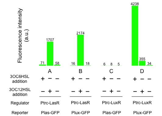
If the GFP expression level of B-2) is as high as that of A-2), it is confirmed that 3OC12HSL-LasR complex crosstalk is occurred.
4. Materials and Methods
4-1. Construction
We used lasR or luxR with constitutive promoter as regulator plasmids, and GFP under las promoter or lux promoter as reporter plasmids. Making pairs from 2 regulators and 2 repressors, we had to prepare 4 plasmid sets. These sets are transformed into different cells (Gray KM et al., 1994).
To construct plasmids in above, we ligated Ptrc-RBS-lasR-TT or Ptrc-RBS-luxR-TT as the regulator, and Plas-RBS-GFP-TT or Plux-RBS-GFP-TT as the reporter plasmid.
Regulator: pSB6A1-Ptrc-lasR / Reporter: pSB3K3-Plas-GFP (JM2.300)…Ptrc-lasR and Plas-GFP cell Regulator: pSB6A1-Ptrc-lasR / Reporter: pSB3K3-Plux-GFP (JM2.300)…Ptrc-lasR and Plux-GFP cell Regulator: pSB6A1-Ptrc-luxR / Reporter: pSB3K3-Plas-GFP (JM2.300)…Ptrc-luxR and Plas-GFP cell Regulator: pSB6A1-Ptrc-luxR / Reporter: pSB3K3-Plux-GFP (JM2.300)…Ptrc-luxR and Plux-GFP cell pSB6A1-Ptet-GFP (JM2.300)…positive control pSB6A1-Promoterless-GFP (JM2.300)…negative control
4-2. Strain
JM2.300
4-3. Protocol
1. O/N -> FC -> Induction
1.1 Prepare overnight culture of each cell (GFP posi, GFP nega, sample) at 37°C for 12 h.
(=> O/N)
1.2 Take 30 microL (from GFP posi, GFP nega, sample) of the overnight culture of inducer cell into LB (3 mL) + antibiotics (Amp 50 microg/mL+ Kan 30 microg/mL).
(=> Fresh Culture)
1.3 Incubate the flesh culture of cells (GFP posi, GFP nega, sample) until the observed OD600 reaches around 0.50.
1.4 Dilute the flesh culture in 1:5 by the following conditions:...LB (3 mL) + antibiotics (Amp 50 microg/mL + Kan 30 microg/mL)
+ 5 microM 3OC6HSL (3 microL)...LB (3 mL) + antibiotics (Amp 50 microg/mL + Kan 30 microg/mL)
+ 5 microM 3OC12HSL (3 microL)...LB (3 mL) + antibiotics (Amp 50 microg/mL + Kan 30 microg/mL)
+ 5 microM DMSO (3 microL)
1.5 Incubate the flesh culture of diluted inducer cell for 4 h at 37°C.
(=> Induction)
2. Measurement (Flow cytometer)
2.1 Measure all samples' OD600.
2.2 Dilute all samples with 1X PBS to keep OD600 in the range from 0.2 to 0.5.
2.3 Take 1 mL (from all samples) into a disposal tube (for flow cytometer).
2.4 Centrifuge them at 9000 g, 4°C, 1 min. and take their supernatant away.
2.5 Suspend all samples with 1 mL 1X PBS.
2.6 Measure all samples.
2.7 Save and organize data.
5. Result of the assay
From above chart (Fig. 3-1-2), it turns out the following. las promoter is activated by 3OC12HSL-LasR complex. Similarly, lux promoter is activated by 3OC12HSL-LasR complex, too. From this result we confirm that crosstalk by 3OC12HSL-LasR complex really happens.
 "
"


