Team:Imperial College/PHB production
From 2013.igem.org
| Line 8: | Line 8: | ||
O/N cultures of MG1655 transformed with either control or phaCAB plasmid were spread onto LB-agar plates with 3% glucose and Nile red staining. | O/N cultures of MG1655 transformed with either control or phaCAB plasmid were spread onto LB-agar plates with 3% glucose and Nile red staining. | ||
{| class="wikitable" style="margin: 1em auto 1em auto;" | {| class="wikitable" style="margin: 1em auto 1em auto;" | ||
| - | [[File: | + | |[[File:EV_vs._phaCAB_red_-_reduced_background.PNG|thumbnail|right|400px|<b>Initial work with plastic synthesis in the native promoter.</b> Nile red staining was used to show expression of the plastic by fluorescence imaging. Control cells with empty vector are shown on the left, while native phaCAB transformed MG1655 is on the right.]] |
|[[File:27-9-13phaCABall.jpg|thumbnail|right|400px|<b>phaCAB P(3HB) synthesis constructs transformed into MG1655</b> Strains were grown on Nile red plates, which stain the PHB strongly and fluoresce in presence of PHB. On the left are MG1655 cells with an empty vector (no fluorescence; no plastic), at the bottom is the native promoter (i.e. low fluorescence, some plastic). At the top and right we have our constitutive and hybrid promoter (respectively), which both show high expression and thus fluoresce very clearly.]] | |[[File:27-9-13phaCABall.jpg|thumbnail|right|400px|<b>phaCAB P(3HB) synthesis constructs transformed into MG1655</b> Strains were grown on Nile red plates, which stain the PHB strongly and fluoresce in presence of PHB. On the left are MG1655 cells with an empty vector (no fluorescence; no plastic), at the bottom is the native promoter (i.e. low fluorescence, some plastic). At the top and right we have our constitutive and hybrid promoter (respectively), which both show high expression and thus fluoresce very clearly.]] | ||
|} | |} | ||
Revision as of 01:11, 1 October 2013
Contents |
PHB production
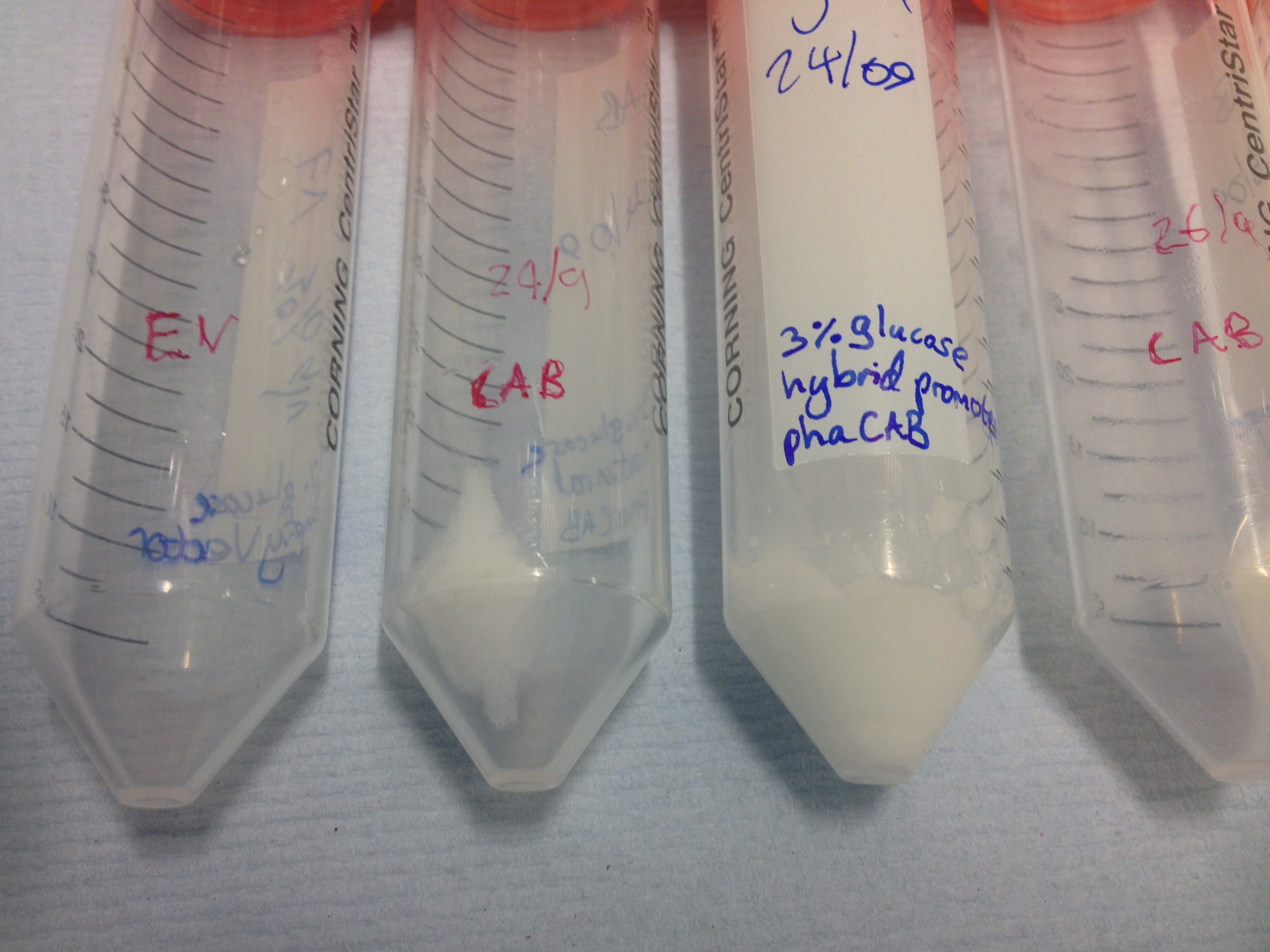
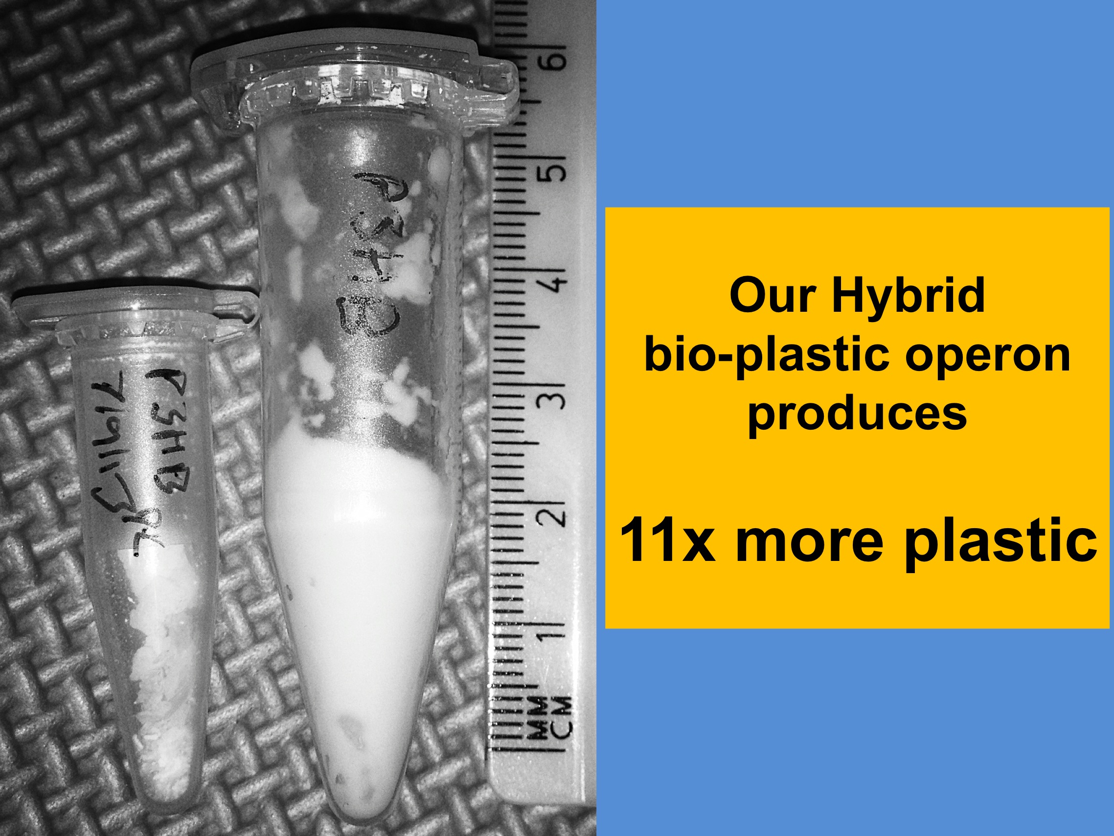
During our project we successfully synthesised the bioplastic P(3HB). We have stained it in the cells on the agar plate and are working hard to do so under the microscope as well. We succesfully extracted PHB from cultures and would also like to detect 3HB monomers from the "white stuff" as an elegant proof of chemical composition.
Nile red staining
O/N cultures of MG1655 transformed with either control or phaCAB plasmid were spread onto LB-agar plates with 3% glucose and Nile red staining.
Conclusion: The red staining indicates the production of P(3HB). More importantly our new Biobricks [http://parts.igem.org/wiki/index.php?title=Part:BBa_K1149051 hybrid promoter phaCAB BBa_K1149051] and [http://parts.igem.org/wiki/index.php?title=Part:BBa_K1149052 constitutive phaCAB BBa_K1149052] produce more P(3HB) than the native phaCAB operon
Extraction of P3HB
We extract P3HB using a technique which first disrupts the cell membranes and then degrades the remaining parts of the cell with bleach. See the protocols section for more details.
Once the cultures have been centrifuged and the supernatant poured off , the biomass is clearly seen as cell pellets.
Purification of P(3HB)
3HB assay
 "
"


