Team:ETH Zurich/Experiments 3
From 2013.igem.org
Contents |
What about those hydrolases ?
We use hydrolases able to make a colorimetric response by converting a specific substrate. We will use 5 hydrolases : Beta-Galactosidase (lacZ),Beta-Glucuronidase (GusA); alkaline phosphatase (phoA);Glycoside hydrolase (NagZ) and the carboxyl esterase (aes).
On this page you will find all relevant informations about the hydrolases and enzyme substrate reaction based on papers and our results.
Enzyme-substrate reactions
We have cloned fluorescent receiver systems as backup for our circuit in case the hydrolase reaction do not work properly.
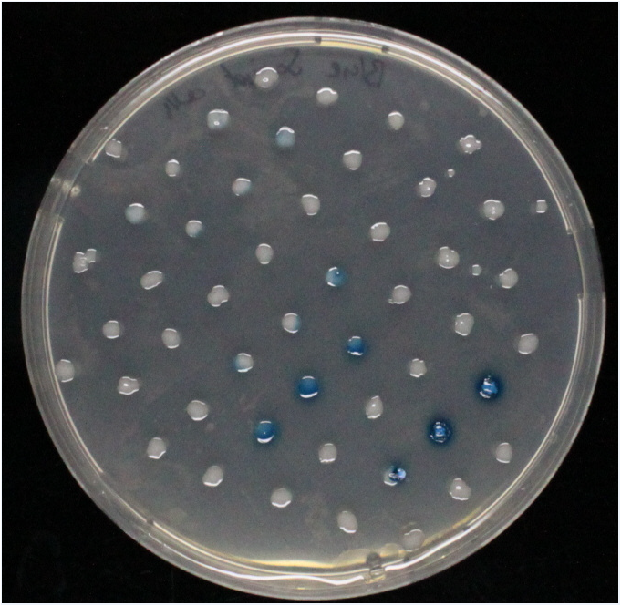
The enzyme substrate reactions take less than 5 minutes and are visible by eye.
Our minesweeper become better and better so keep on track for updates !
| Hydrolase | Complementary substrate / IUPAC name | Visible color | Concentration[M] | Time(s)for colorimetric response | ||
|---|---|---|---|---|---|---|
| LacZ | Beta-Galactosidase | X-Gal | 5-Bromo-4chloro-3-indolyl-beta-galactopyranoside | Blue | ||
| LacZ | Beta-Galactosidase | Green-beta-D-Gal | N-Methyl-3-indolyl-beta-D_galactopyranoside | Green | ||
| GusA | Beta-glucuronidase | Magenta glucuronide | 6-chloro-3-indolyl-beta-D-glucuronide-cycloheylammonium salt | Red | ||
| PhoA | Alkaline phosphatase | pNPP | 4-Nitrophenylphosphatedi(tris) salt | Yellow | ||
| Aes | Carboxyl esterase | Magenta butyrate | 5-bromo-6-chloro-3-indoxyl butyrate | Magenta | ||
| NagZ | Glycoside hydrolase | X-glucosaminide X-Glunac | 5-bromo-4-chloro-3-indolyl-N-acetyl-beta-D-glucosaminide | Blue | ||
What about the hydolases ? How do they work and where do they come from ? Why do we use hydrolases ?
Beta-galactosidase (LacZ)
Alkaline phosphatase(PhoA)
Alkaline phosphatases are used as reporter enzymes in different assays such as Western Blotting and in situ hybridization[1]. Testing human blood for Alkaline Phosphatase levels is a routine test that can reveal different conditions[2].
Alkaline phosphatases cleave phosphate groups from organic compounds by hydrolysis while retaining stereochemistry[3].Alkaline phosphatases, respectively their serum levels, are also related to several diseases e.g. metabolic myopathies and Paget Disease. [4]
The explanation of the mechanism was found on the [http://chemwiki.ucdavis.edu/Organic_Chemistry/Organic_Chemistry_With_a_Biological_Emphasis/Chapter_10%3A_Phosphoryl_transfer_reactions/Section_10.3%3A_Hydrolysis_of__phosphates Chem Wiki ].
While kinase enzymes catalyze the phosphorylation of organic compounds, enzymes called phosphatases catalyze dephosphorylation reactions. Serine phosphatase, for example, catalyzes the following dephosphorylation of phosphoserine residues:
[[File:]
The reactions catalyzed by kinases and phosphatases are not the reverse of one another: kinases transfer phosphoryl groups from ATP (or sometimes other nucleoside triphosphates) to various organic compounds, while phosphatases transfer phosphoryl groups from organic compounds to water, which is a hydrolysis reaction.
Look again the serine phosphatase reaction. Two very different things could be happening, given the products that result. The reaction could be a phosphoryl transfer reaction, in which a phosphorus-oxygen bond is broken (reaction A). Alternatively, the water could be attacking the carbon of the serine side chain, breaking a carbon-oxygen bond and expelling the phosphate group in a nucleophilic substitution reaction (reaction B).
[[File:]]
In order to find out which of these two mechanisms applies to phosphatases, scientists incubated a phosphatase enzyme with H218O, then used mass spectrometry to determine that the 18O ended up exclusively in the free phosphate (J. Biol. Chem. 1961 236, 2284). This result supports the idea that phosphatase reactions are phosphoryl transfer reactions (reaction A above), not nucleophilic substitutions.
Given this information, we would expect that the phosphatase reactions result in inversion of stereochemistry at the phosphate phosphorus. Experiments with chiral, 17O and 18O-labeled phosphate ester substrates show that this is indeed the case with many phosphatases (J. Am. Chem. Soc. 1993, 115, 2974). In other enzymes, however, dephosphorylation appears to occur with retention of configuration. Glucose-6-phosphate phosphatase is one such example (Biochem. J. 1982, 201, 665).
[[File:]]
How could a phosphoryl transfer reaction result in retention of stereochemistry? Think back to the retaining glycosidase reaction (section 9.2), in which a nucleophilic substitution with overall retention of configuration was achieved via a double displacement mechanism. A nucleophile on the enzyme itself carries out the first nucleophilic attack (step 1) , then is subsequently displaced by water (step 2).
[[File:]]
Could a similar thing be happening here? In order to find out, researchers ran the reaction with radioactive 32P-labeled glucose-6-phosphate, then stopped the reaction midstream in order to try to isolate the predicted enzyme-phosphate intermediate. The enzyme was chopped up into small pieces using proteases (we'll learn about these protein-chopping enzymes in chapter 12), and a radioactive phosphate was found to be covalently attached to an active site histidine (Biochem Biophys Acta 1972, 268, 698). Apparently, this histidine is acting in the role of the 'X' group in the figure above.
Carboxyl esterase (Aes)
Glycoside hydrolase (NagZ)
Beta-glucuronidase (GusA)
Do the substrates and enzymes cross-react ?
An enzyme-substrate test matrix (Figure 6) was established to test each substrate against each enzyme. The results were as expected (Figure 6.2) and no cross reaction is visible. The NagZ-X - glucosaminide X-Glunac reveal some difficulties in the liquid culture as well as on the agar plate.
 "
"


