Team:TU-Eindhoven/Background
From 2013.igem.org


Contents |
Background
In general terms, diagnostic imaging is proving to be a crucial aid in the medical practice. A broad range of diseases are diagnosed in an earlier, faster and easier manner due to the significant advances in the field of diagnostics. MRI is one of the most important diagnostic imaging systems. It provides doctors and researchers with the means for acquiring high quality 3D images of a patient's body. Hence, MRI has become an irreplaceable tool in the detection of cancers.
MRI relies on measuring magnetization of magnetic nuclei subjected to radio-frequency irradiation inside a magnetic field. BulteRepGenesA.A. Gilad, K. Ziv, M.T. McMahon, P.C.M. van Zijl, M. Neeman, and J.W.M. Bulte, MRI Reporter Genes. Nuclear Medicine 49, 1905-1908 (2008) There are many nuclei that can be studied and considered, the hydrogen nucleus being the most common one. MRI detects the properties of the water signal, i.e. the spin density, its relaxation back to equilibrium after excitation, and its signal augmentation. Any tissue alteration in terms of these three properties provides contrast. Aside from water, magnetic resonance spectroscopy, better known as MRS, can identify individual proton signals in metabolites and nuclei different from protons. The signal obtained from diverse nuclei depends on the abundance of it in the tissue of interest and the chemical interaction between the nuclei and other molecules present in the tissue. The latter allows the development of different contrasts by performing several manipulations with a variety of MRI acquisition schemes.
Contrast agents are used worldwide in MRI for signal enhancement. Using them permits a greater differentiation between healthy and damaged tissue, moreover it allows a better visualization of different structures. Nowadays, the most common clinically used agents are complexes of Gd3+ ions that shorten the relaxation time of free water protons. LenkinskiCESTBasicToAppsE. Vinogradov, A. Dean Sherry, and R.E. Lenkinski, CEST: From basic principles to applications, challenges and opportunities. Magnetic Resonance , 1-18 (2012) These agents are non-selective, and have a uniform distribution within the extracellular space after intravenous injection. LaufferGdChelatesP. Caravan, J.J. Ellison, T.J. McMurry, and R.B. Lauffer, Gadolinium(III) chelates as MRI contrast agents: structure, dynamics, and application. Chemical Reviews 99, 2293-2352 (1999) As previously mentioned, there are many other contrast techniques that are based on the intrinsic properties of tissue, i.e. coupling to neighboring nuclei, chemical exchange or flow. GrahamMTMRIR.M Henkelman, G.J. Stanisz, and S.J. Graham, Magnetization transfer in MRI: a review. NMR Biomed. 14, 57-64 (2001) In the 1990's, Balaban and colleagues introduced a new class of contrast agents for MRI known as Chemical Exchange Saturation Transfer contrast agents.
CEST 101: What is it and how does it work?

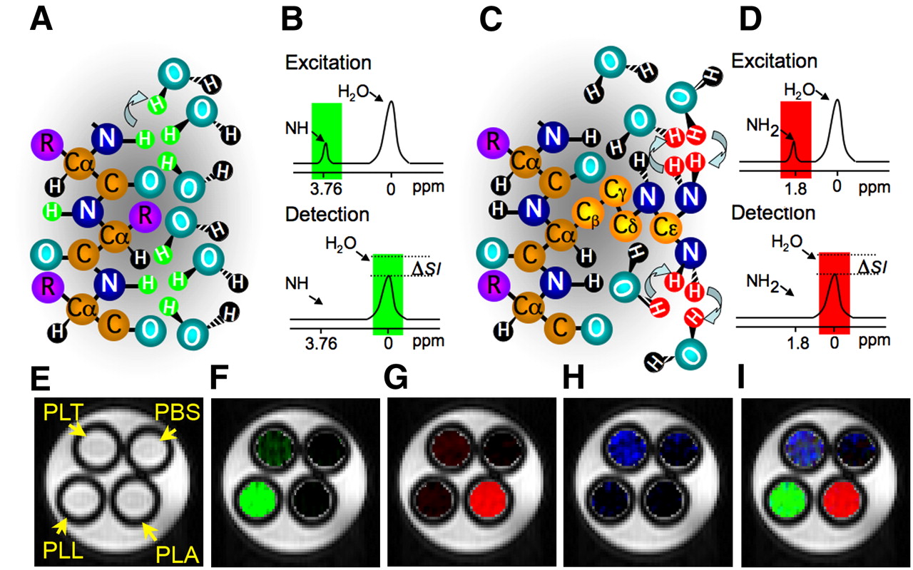
This type of MRI contrast relies on the chemical exchange of protons of solutes such as contrast agents with bulk water. Many organic and organometallic compounds can be excited in a selective manner and detected sensitively due to the fact that they contain enough protons with a proper chemical exchange rate and specific MRI frequencies. A saturation pulse, which is a radio frequency pulse, is applied at the resonance frequency of the exchangeable proton. The signal loss originated from it is transferred through the exchange to bulk water, creating a fractional reduction in the water signal in this manner. WoodChemExSTContrastAgentsA.D Sherry, and M. Woods, Chemical exchange saturation transfer contrast agents for magnetic resonance imaging. Annu Rev Biomed Eng. 10, 391-411 (2008) Some of the molecules that have been proven to work as potential agents are small diamagnetic molecules, complexes of paramagnetic ions, endogenous macromolecules, dimers and liposomes.LenkinskiCESTBasicToApps
CEST contrast agents have two main advantages over other types of contrast agents. First, they can be turned on and off as a consequence of the contrast being detectable only when the saturation pulse is applied at a very specific frequency. In other words, the contrast agent is switchable, going from visible to invisible according to the frequency at which the pulse is applied. The second advantage is that CEST contrast agents allow the imaging simultaneously of more than one target, using different contrast agents with different excitation frequencies. BulteRepGenes
Why use bacteria as production factory and delivery system?
Bacteria can sense their environment, differentiate between cell types, and deliver proteins to eukaryotic cells.VoigtControlledInvasionCancerCellsJ.C. Anderson, E.J. Clarke, A.P. Arkin, and C.A. Voigt, Environmentally Controlled Invasion of Cancer Cells by Engineered Bacteria. Molecular Biology 355, 619-627 (2006). A rapidly evolving research area in the field of oncology is the one focused on bacterial based cancer therapies. In this type of therapies, bacteria like E. Coli and Salmonella typhimurium are used as tumor targeting systems. These bacteria are used to deliver cytotoxic agents, immunostimulatory cytokine, and genetic material to enable host-cell production of these molecules, and induce cell death in this way. forbesBactN.S. Forbes, Engineering the perfect(bacterial) cancer therapy. Nature , 785-794 (2010) Tumors present a hypoxic environment, and thus they can be targeted by bacteria. Hypoxic conditions can also function as a trigger for the production of specific proteins, in our case CEST proteins. Generating the CEST proteins only under hypoxic circumstances ensures that the contrast is created only in the tumor region, consequently working as a tumor targeting system for drug delivery at the same time.
This type of approach could prove to be safer, more efficient, and less expensive due to its biospecificity, or ability to localize and target a specific site.GaoCureCancerK. Gao, E. coli: The Cure for Cancer?. The Triple Helix , 12-13 (2010) Moreover, the use of bacteria as a vector to generate the CEST MRI contrast agent in situ allows higher carrying capability than conventional methods.
There is a wide range of possible applications, being the most logical one the usage of the E.coli based CEST contrast agent for MR imaging in humans with diverse types of cancer. Other applications might be the study of bacterial infections and drug testing.
Of course there is always the safety issue question when thinking about introducing bacteria into the bloodstream. This issue can be adjourned by disguising the bacteria, commanding the E. coli to grow an alternate carbohydrate surface layer instead of chains which would prevent an immune response from happening.GaoCureCancer Circumventing the immune response issue, the bacteria are able to carry out their tasks.
References
 "
"


