Team:Linkoping Sweden/Modeling
From 2013.igem.org
(Difference between revisions)
| Line 16: | Line 16: | ||
[[File:ljusmodel_illustration.png]] | [[File:ljusmodel_illustration.png]] | ||
| + | |||
This picture describes how the model will explain the antigen/antibody-complex binding and the light difference. | This picture describes how the model will explain the antigen/antibody-complex binding and the light difference. | ||
| + | As antibody is added to the sample (T=0 min) containing the unknown amount of HEWL-antigen green light is emitted from luciferase. When the RFP-antigen is added (T=10 min) the decrease in green fluorescence and the increase in red fluorescence will be detectable and the detector will thus give a signal saying if it is HEWL-antigen in the sample or not. | ||
[[File:Ljusmodel.png]] | [[File:Ljusmodel.png]] | ||
{{Template:Team Linkoping footer}} | {{Template:Team Linkoping footer}} | ||
Revision as of 13:32, 19 August 2013


System Modeller light sensitivity model
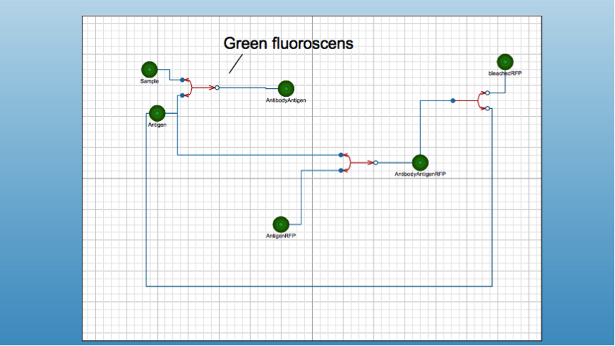
This is the model that describes the detection of light output from our designed antibody/antigen complex. The model-program vill be incorporated into the physical biosensor to determine the amount of antigen in the sample.
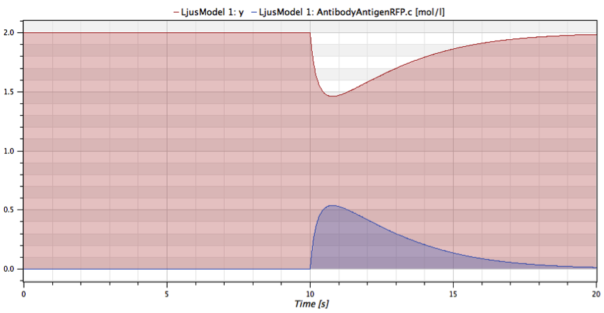
This picture describes how the model will explain the antigen/antibody-complex binding and the light difference.
As antibody is added to the sample (T=0 min) containing the unknown amount of HEWL-antigen green light is emitted from luciferase. When the RFP-antigen is added (T=10 min) the decrease in green fluorescence and the increase in red fluorescence will be detectable and the detector will thus give a signal saying if it is HEWL-antigen in the sample or not.


 "
"
