Team:NYMU-Taipei/Experiment/Wet Lab
From 2013.igem.org
Mastershot (Talk | contribs) |
|||
| Line 1: | Line 1: | ||
{{:Team:NYMU-Taipei/Header}} | {{:Team:NYMU-Taipei/Header}} | ||
| - | ==Coding Sequence Testing | + | =N. ceranae experiment protocols= |
| + | ==Extraction of N. ceranae Spores from Bees== | ||
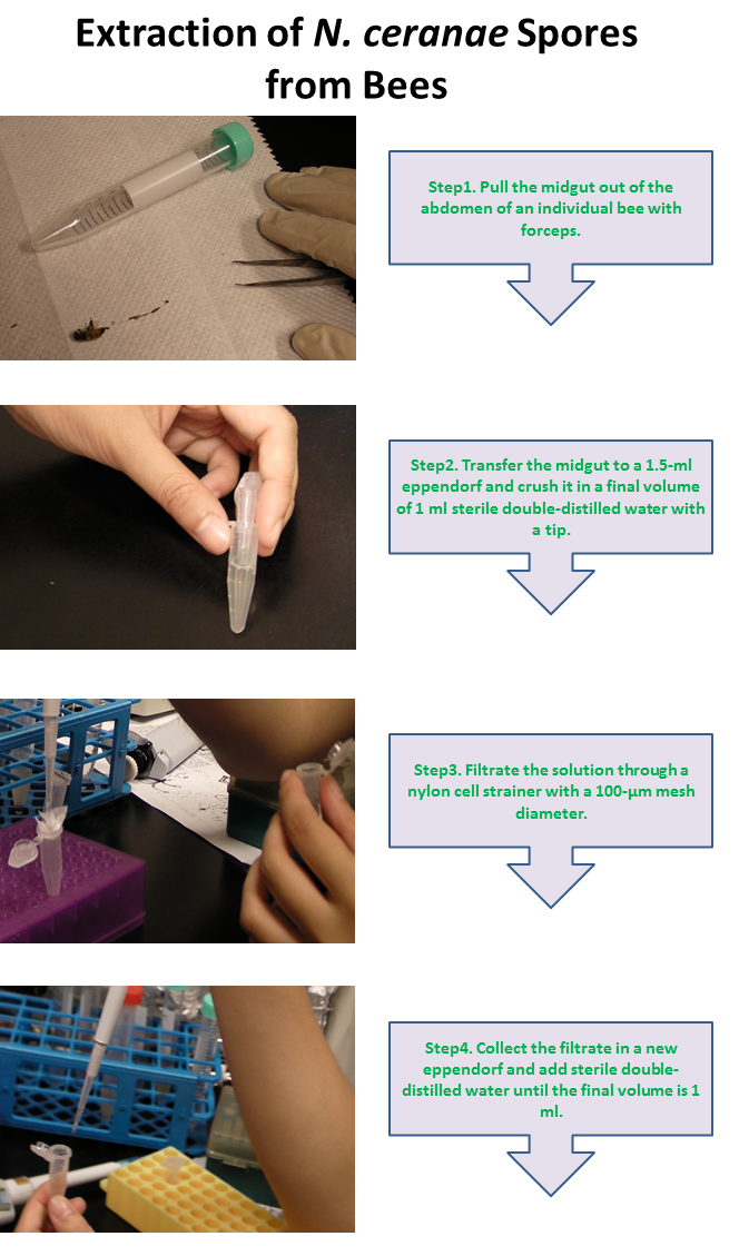
| + | [[File:Extraction.png|frame|center]] | ||
| + | ==Purification of N. ceranae Spores== | ||
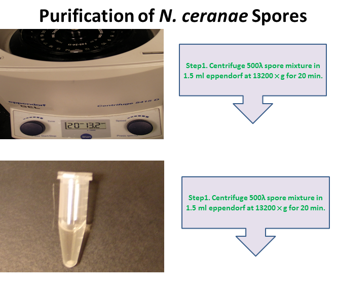
| + | [[File:Nymu-Purification.png|frame|center]] | ||
| + | ==N. ceranae Genomic DNA Extraction== | ||
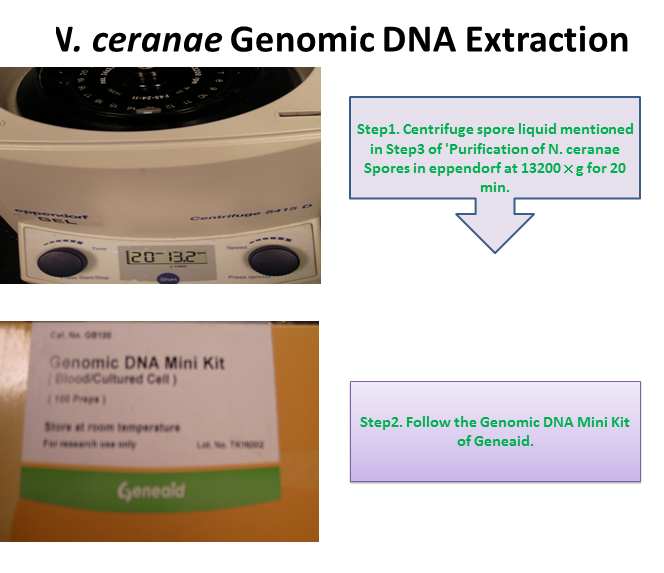
| + | [[File:Nymu-Genomic.png|frame|center]] | ||
| + | ==Germination of N. ceranae Spores== | ||
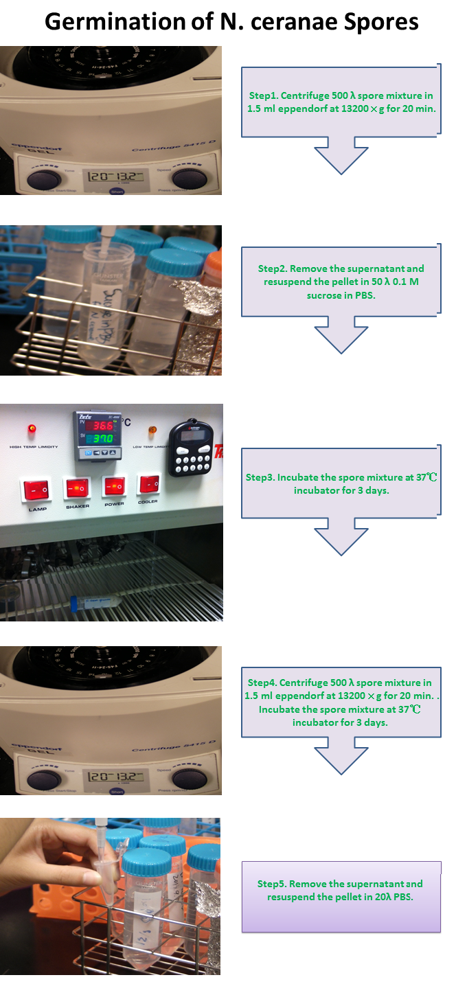
| + | [[File:Nymu-Germination.PNG|frame|center]] | ||
| + | |||
| + | =Coding Sequence Testing= | ||
We define the germination degree as follows: | We define the germination degree as follows: | ||
{| | {| | ||
| Line 11: | Line 21: | ||
|} | |} | ||
| - | + | ==Novel protocol for differentiation of N. apis and N. ceranae== | |
| - | ==N. ceranae | + | ''Nosema apis'' and ''Nosema ceranae'' are two genetically related microsporidian pathogens of the western honey bee or European honey bee (''Apis mellifera''). Both of them have similar life cycles, shorten the adult bee’s lifespan and impact honey production, but only ''N. ceranae'' is suspected to induce the CCD problem. Also, ''N. apis'' is well controlled by the use of fumagillin while ''N. ceranae'' escapes the antimicrobial agent. (http://www.plospathogens.org/article/info%3Adoi%2F10.1371%2Fjournal.ppat.1003185) In recent years, ''N. ceranae'', originally found in the Asian honey bee (''Apis cerana''), appears to have become more dominant than ''N. apis'' in the western honey bees. The distinction between ''N. apis'' and ''N. ceranae'' is thus important and still worth further research. |
| - | + | Light microscopy method based on morphology revealed that spores of ''N. ceranae'' were oval or rod shaped like a rice, and were slightly thinner than ''N. apis''. | |
| - | + | ||
| - | + | ||
| - | + | ||
| - | + | ||
| - | + | ||
| - | + | ||
| - | + | ||
| - | + | ||
| - | + | ||
| - | + | ||
| - | + | ||
{{:Team:NYMU-Taipei/Footer}} | {{:Team:NYMU-Taipei/Footer}} | ||
Revision as of 13:58, 27 September 2013


Contents |
N. ceranae experiment protocols
Extraction of N. ceranae Spores from Bees
Purification of N. ceranae Spores
N. ceranae Genomic DNA Extraction
Germination of N. ceranae Spores
Coding Sequence Testing
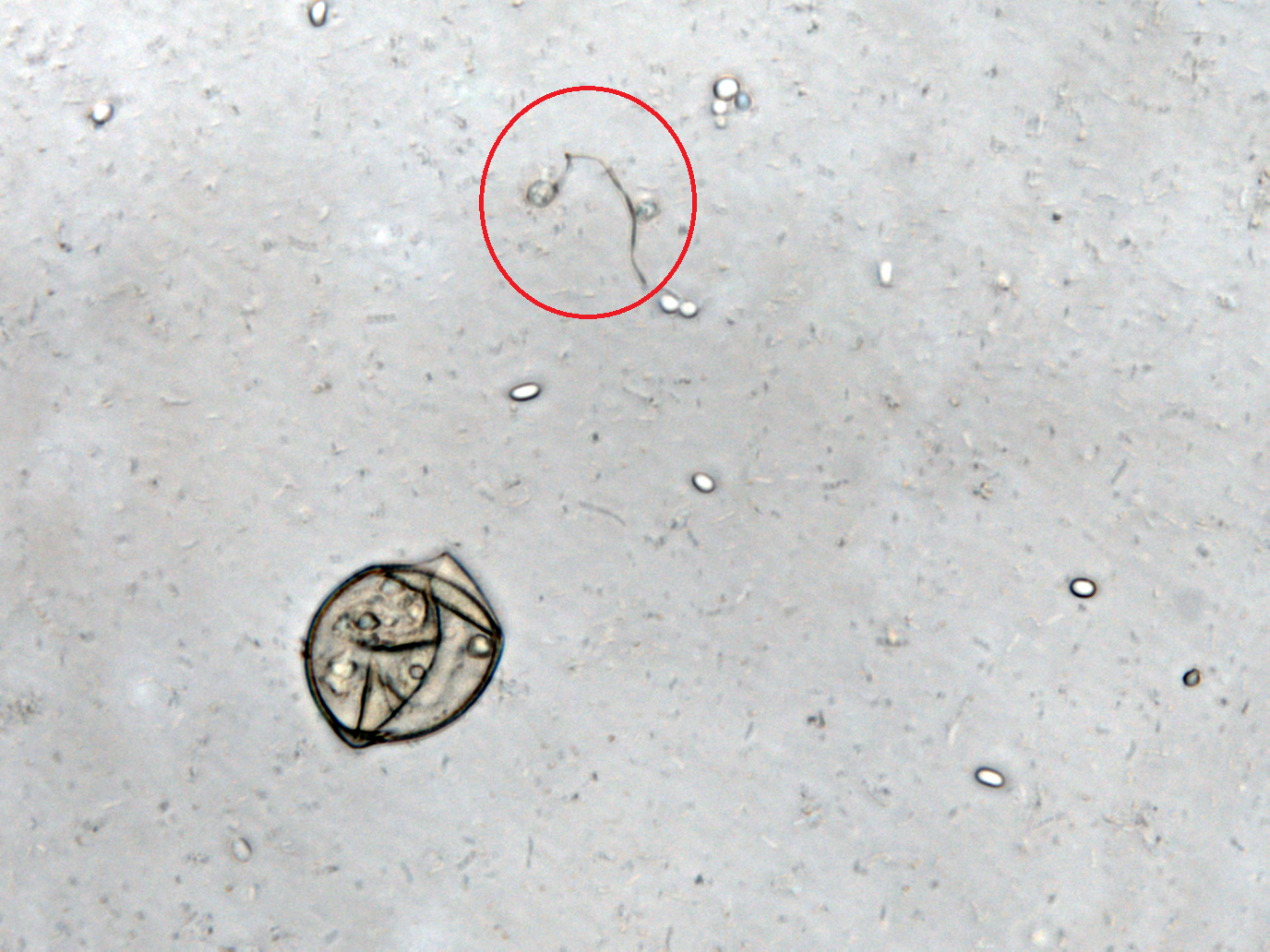
We define the germination degree as follows:
Novel protocol for differentiation of N. apis and N. ceranae
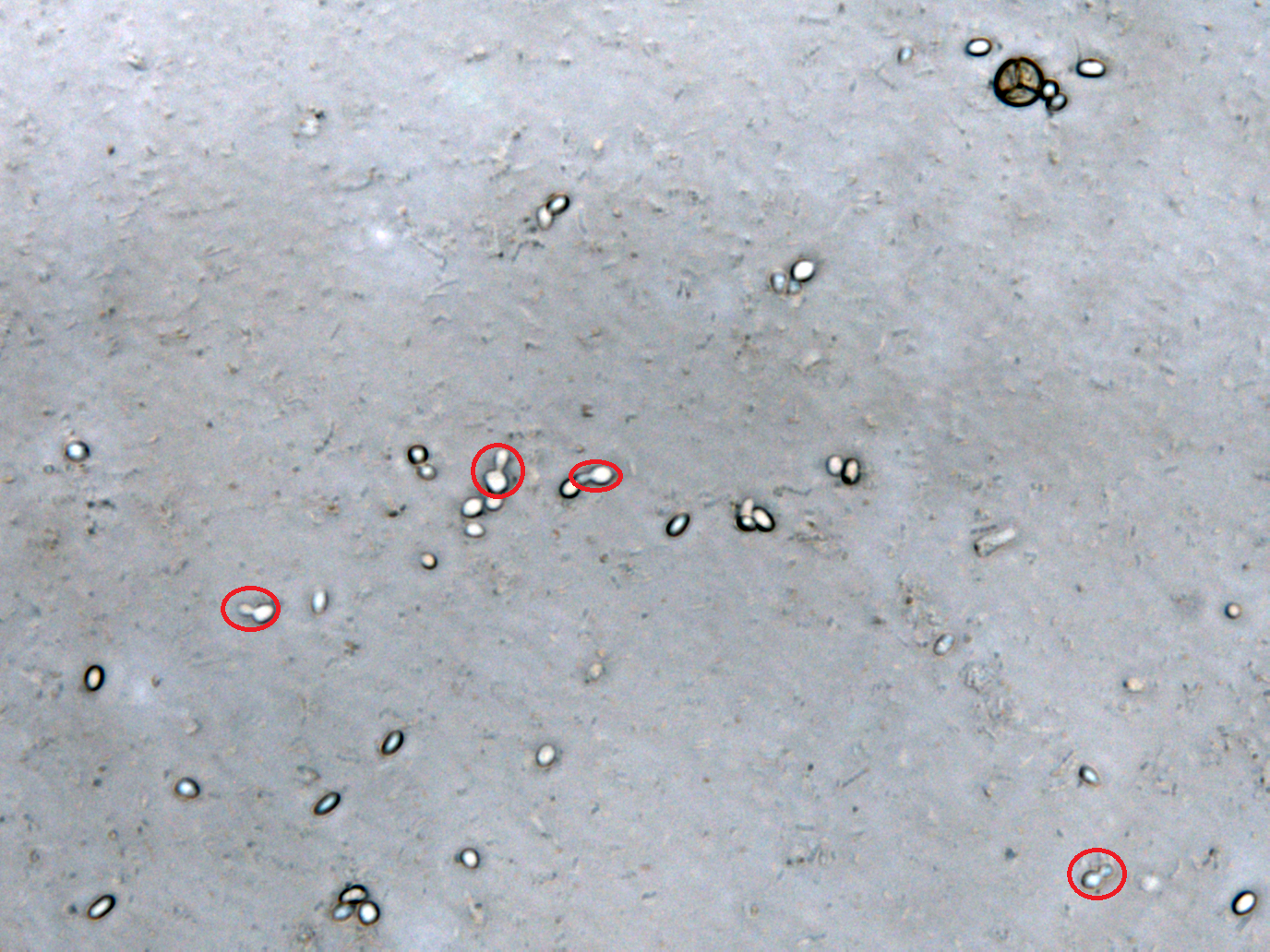
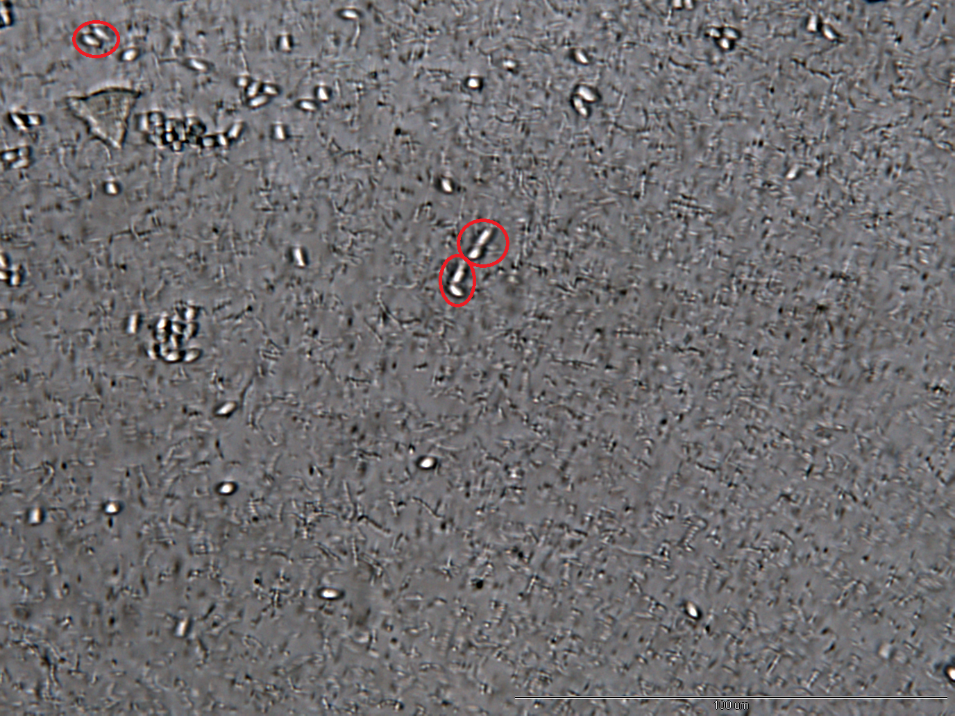
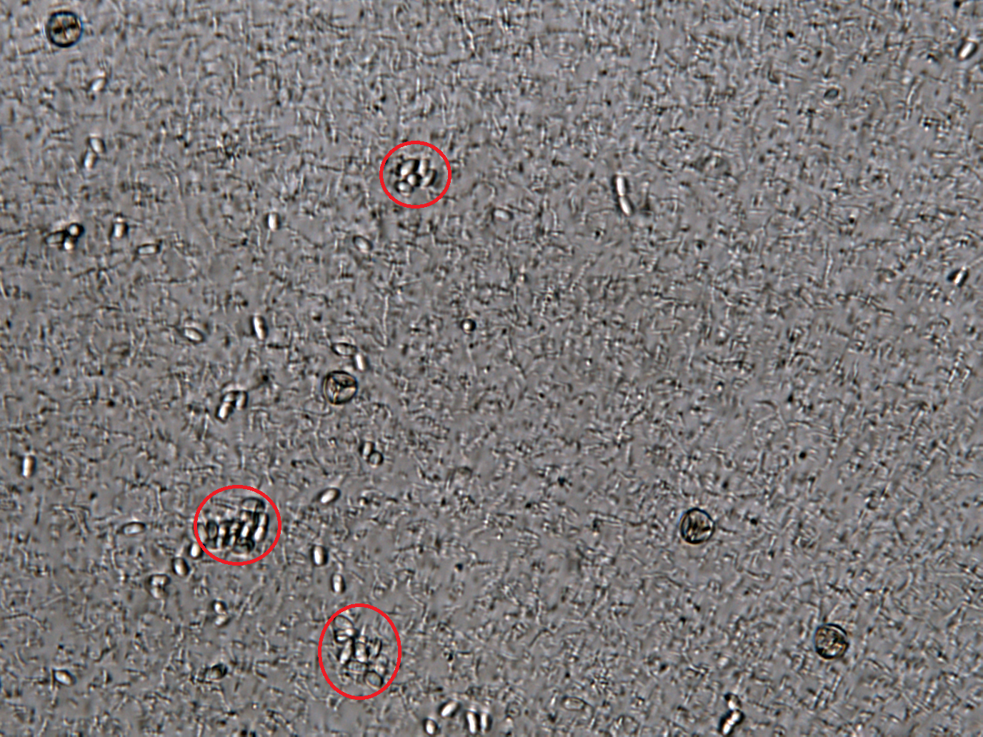
Nosema apis and Nosema ceranae are two genetically related microsporidian pathogens of the western honey bee or European honey bee (Apis mellifera). Both of them have similar life cycles, shorten the adult bee’s lifespan and impact honey production, but only N. ceranae is suspected to induce the CCD problem. Also, N. apis is well controlled by the use of fumagillin while N. ceranae escapes the antimicrobial agent. (http://www.plospathogens.org/article/info%3Adoi%2F10.1371%2Fjournal.ppat.1003185) In recent years, N. ceranae, originally found in the Asian honey bee (Apis cerana), appears to have become more dominant than N. apis in the western honey bees. The distinction between N. apis and N. ceranae is thus important and still worth further research. Light microscopy method based on morphology revealed that spores of N. ceranae were oval or rod shaped like a rice, and were slightly thinner than N. apis.
 "
"


