 "
"
Team:TU-Eindhoven/CESTAgentTesting
From 2013.igem.org
Pascalaldo (Talk | contribs) (→Abstract) |
Pascalaldo (Talk | contribs) (→Abstract) |
||
| Line 3: | Line 3: | ||
=CEST Agent Testing= | =CEST Agent Testing= | ||
=== Abstract === | === Abstract === | ||
| - | Until recently, the most common way of generating MRI contrast was to use heavy metal based contrast agents. Examples of these are the commercially available Gadolinium(III) based Magnevist and the Iron Oxide based Resovist. | + | Until recently, the most common way of generating MRI contrast was to use heavy metal based contrast agents. Examples of these are the commercially available Gadolinium(III) based Magnevist and the Iron Oxide based Resovist. A downside of using heavy metals is the toxicity of the agents in the human body{{:Team:TU-Eindhoven/Template:Ref | id=GNSF | author=T Grobner and FC Prischl | title=Gadolinium and nephrogenic systemic fibrosis| journal=Kidney International | edition=72 | pages=260–264 | year=2007 }}{{:Team:TU-Eindhoven/Template:Ref | id=Gsrf34 | author=Ihsan Ergün, Kenan Keven, et all. | title=The safety of gadolinium in patients with stage 3 and 4 renal failure | journal=Nephrology Dialysis Transplantation | edition=21 | pages=697-700 | year=2006 }}{{:Team:TU-Eindhoven/Template:Ref | id=Gint | author=I. Buhaescu and H. Izzedine | title=Gadolinium-induced nephrotoxicity | journal=The International Journal of Clinical Practice | edition=62 | pages=1113-1118 | year=2008 }}. Fortunately, a new type of MRI has been developed, which allows for the use of organic compounds as contrast agent. We chose several Arginine and Lysine rich proteins for testing them for CEST contrast. Hereto the proteins were expressed aerobically in BL21 bacteria. The bacterial cell pellets were fixed using PFA and then tested in a 7 T Bruker MRI machine. From these experiments it was concluded that the most promising candidate for Lysine based CEST contrast agents is 1PJN, of which the lysine peak was clearly distinguishable from the rest of the signal. No clear results have been achieved for Arginine based CEST contrast agents. |
=== Introduction === | === Introduction === | ||
Revision as of 23:26, 4 October 2013


Contents |
CEST Agent Testing
Abstract
Until recently, the most common way of generating MRI contrast was to use heavy metal based contrast agents. Examples of these are the commercially available Gadolinium(III) based Magnevist and the Iron Oxide based Resovist. A downside of using heavy metals is the toxicity of the agents in the human bodyGNSFT Grobner and FC Prischl, Gadolinium and nephrogenic systemic fibrosis. Kidney International 72, 260–264 (2007)Gsrf34Ihsan Ergün, Kenan Keven, et all., The safety of gadolinium in patients with stage 3 and 4 renal failure. Nephrology Dialysis Transplantation 21, 697-700 (2006)GintI. Buhaescu and H. Izzedine, Gadolinium-induced nephrotoxicity. The International Journal of Clinical Practice 62, 1113-1118 (2008). Fortunately, a new type of MRI has been developed, which allows for the use of organic compounds as contrast agent. We chose several Arginine and Lysine rich proteins for testing them for CEST contrast. Hereto the proteins were expressed aerobically in BL21 bacteria. The bacterial cell pellets were fixed using PFA and then tested in a 7 T Bruker MRI machine. From these experiments it was concluded that the most promising candidate for Lysine based CEST contrast agents is 1PJN, of which the lysine peak was clearly distinguishable from the rest of the signal. No clear results have been achieved for Arginine based CEST contrast agents.
Introduction
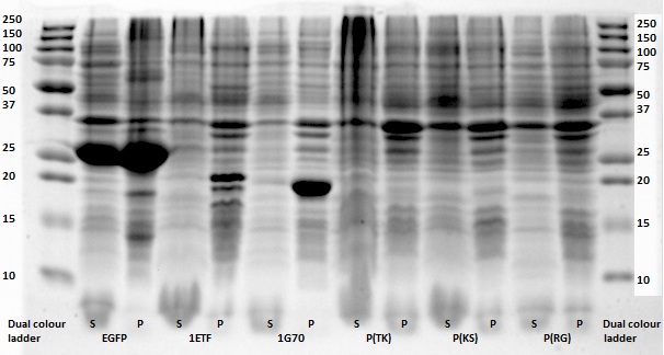
Initially, a total of 10 constructs with proteins were designed, all containing a high percentage of lysine and/or arginine, which have a chemical shift which can be used for CEST imaging. Four of them were naturally occurring proteins, native to either E. coli or to other organisms, in which case the protein sequence was optimized to match codon prevalence in E. coli. These were 1G70, 1ETF, 1PJN Chain-1 and Human Protamine 1 (HPM1, optimized). The other five were non-natural polypeptides consisting of a two repeating dipeptides, namely poly(arginine-glycine) (P(RG)), poly(lysine-serine) (P(KS)), poly(arginine-serine) (P(RS))and poly(threonine-lysine) (P(TK)). Like HPM1, these we E. coli optimised. Additionally, an EGFP construct would also be inserted and expressed as a positive control.
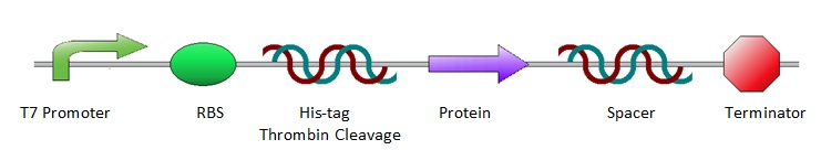
All the protein sequences were cut out of the ordered pUC57(s) vectors by NheI and XhoI restriction enzymes and ligated into pET28a vectors. As a result, all constructs shared the same lay-out, starting with prescribed iGEM restriction sites EcoRI, NotI and XbaI, followed by an T7 promotor site and ribosome binding site. Hereafter are a poly-histidine tag (for protein purification), thrombin cleavage site and NheI restriction site (to isolate the protein sequence for ligation into a pET28a vector). Subsequently comes the variable part, the protein coding sequence. Then follow an XhoI restriction site and random spacer sequence, positioning the terminator at 30 base pairs from the stop codon. After the terminator come three prescribed iGEM end restriction sites, SpeI, NotI, PstI, plus a HindIII restriction site.
Methods and Results
Cloning
Once the pUC57(s) constructs were delivered, we could transform them into NB bacteria for amplification of the vectors. When they were amplified, a digestion on the constructs as well as on the pET28a vector is performed with NheI and XhoI restriction enzymes to cut out just the protein sequence out of the pUC57(s) construct. Chosen was for pET28a since it has a promoter that can be induced by IPTG which is helpful and on the otherhand there was much DNA left in stock. Once the pUC57(s) constructs as well as pET28a vector were digested a gel extraction was performed to cut out the desired bands. Then a ligation is performed to get the protein sequence from the pUC57(s) constructs into the pET28a vector. After ligation the constructs were plated with kanamycin antibiotics and grown overnight to create colonies. To check whether ligation was succeeded a colony PCR was performed. Primers used in the ligation of pET28a vectors were the T7 Forward and T7 Reverse (terminator) primers.
After completing the PCR protocol, an agarose gel electrophoresis was executed on the products and visualised using UV spectrometry to see whether the ligation was successful or not.
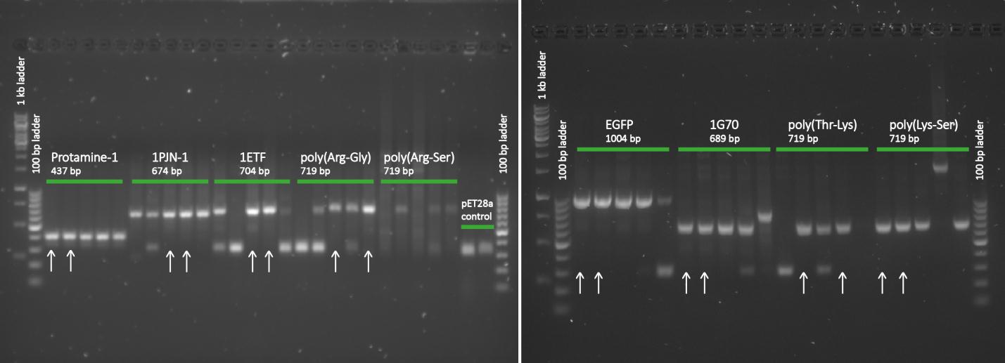
Except for the poly(arginine-serine), all constructs yielded successfully ligated products for further processing. For each protein sequence, two of these were selected based on the intensity and the accurate weights of the appearing bands. Then from the chosen constructs, small cultures were set in, again with kanamycin, and grown overnight. To retain the vectors, a miniprep procedure was performed and once the constructs were sequenced and seemed to be correct, they were transformed into BL21 bacteria for protein expression.
Protein Expression and Sampling for CEST Measurements
Large cultures with the BL21 bacteria, containing our constructs, were set in and were grown until the optical density was about 0.6. Then protein expression was induced by adding IPTG to induce the promoter and protein expression was then performed overnight at 37°C.
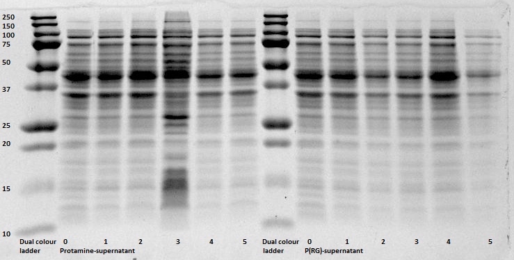
For protein retention a Bugbuster protocol was performed. Once pellets had formed during the Bugbuster protocol, samples of the pellets were taken for CEST measurements. This was done by taking about 300-400 mg of the pellet (this was done to make comparison of the CEST measurements possible) and the pellets were placed into a PCR tube and the cells were fixated with 4% PFA (paraformaldehyde) and placed in the refrigerator at 4°C for at least two days.

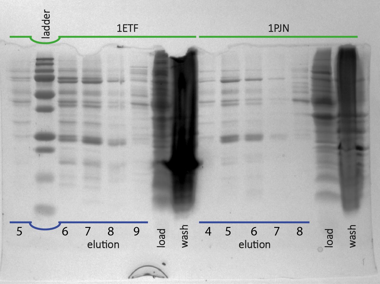
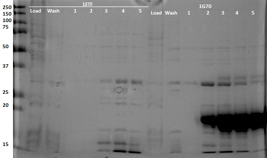
After expression of 1ETF and 1PJN, a Nickel-column purification was performed (for all proteins) to purify the proteins (since they have an His-tag). Samples were taken from each step (load, wash and elution) for further analysis (SDS gel analysis and Bradford assay), after rebuffering to TRIS buffer, since imidazole can cause protein denaturation.
MRI Measurements
The entire pellet (bacteria containing our proteins) was then measured in a 7 T Bruker MRI machine. First, the correct water frequency was determined, the machine was shimmed, i.e. a homogeneous magnetic field was created. The first measurement was a T2 weighed image for general orientation. Subsequently local shimming was performed on each of the separate pellets. For the final measurements, the saturation pulse was set to vary from $\pm -4 ppm$ to $\pm +4 ppm$ (relative to water), the measurements were averaged over 8 separate scans. Also a $S_{0}$ (without saturation pulse) image was taken. For more information about the theory of CEST Imaging, see MRI Processing.
Discussion and Conclusions
All in all, the most promising candidate agent seems to be 1PJN (BBa_K1123015). The lysine peak of this agent is clearly distinguishable from the rest of the signal. As a arginine based compound, no clear results have been achieved and therefore no recommendation can be given, other than that the arginine based agents are less suitable for generating contrast in general. It is therefore our recommendation to use lysine based agents, such as 1PJN. Only for multi-color MRI or imaging lysine rich areas can arginine agents be useful. Another conclusion that can be drawn from the MRI results, is that Poly(KS) has actually been expressed in the bacteria. This is contradictory to the other lab results, where no traces of Poly(KS) on SDS-Page gels were found. The most obvious explanation for these results, is that only a very small part of the Poly(KS) chain was created, which is to be expected due to the largely repetitive and non-optimal DNA of the construct.