 "
"
Team:USTC CHINA/Project/Results
From 2013.igem.org

Stage One:Basic Experiments
Introduction
As the construction and concentration of recombinant plasmid rely heavily on E.coli system in current molecule experiment protocols of B.subtilis, B.subtilis acts only as the secretory expression vector. Therefore, to verify the practicality of our locus and the transdermal function of recombinant transdermal protein on top of its original functions, we conducted basic experiments on E.coli to verify our assumptions.
The following figure shows the stages of our basic experiments:

Results
1. Verifying the Validity of Our Circuit by GFP
E.coli BL21 proved that the standard transdermal locus does work with GFP.

Using GFP to prove the validity of a newly designed circuit is a classical way to verify the expressing of this circuit. As expressions in E.coli involve neither secretory nor sequential problems, we hoped to verify the practicality of our locus by the expression of TD1-GFP. Thus we selected pET22b, which is a common recombinant vector for plasmid construction, as our recombinant vector and E.coli BL21 as engineered bacteria. We fused sequence TD1-GFP with T7 promoter from pET22b downstream and succeeded in expressing fusion protein TD1-GFP.
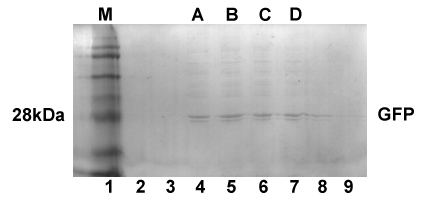 Fig1. SDS PAGE shows the molecule weight of TD1-GFP
2. TD1 Fusion Protein Expression
The expression of recombinant antigen and adjuvant in E.coli BL21.

The practicality of our circus afforded by TD1-GFP enabled our to express recombinant antigen TD1-HBsAg and recombinant adjuvant TD1-LTB successfully. So far our basic molecule experiments have ended with perfection.


3. Verifying the Antigenicity of TD1-Antigen by ELISA
The antigenicity of the TD1-antigen(fusion protein)strongly proved our theoretical basis. We have set 3 different negative control groups(GFP sample, HBsAg transdermal result, normal saline) and one positive group standard HBsAg sample.

4. Transdermal Experiment
TD1-HBsAg is able to penetrate the skin and keep its antigenicity:

To prove that TD1-HBsAg is able to pass across the skin and keep its antigenicity, we utilized a special device in the picture below, whose upper tube and lower tube can be separated by fresh skin peeled from mice, and all we were required to do was fastening the device, adding protein solution to the upper tube and extracting appropriate volume of liquid from the physiological saline in the lower tube to check the concentration of TD1-HBsAg which had crossed the skin during diverse periods.
