Team:ETH Zurich/Experiments 3
From 2013.igem.org
(Difference between revisions)
| Line 62: | Line 62: | ||
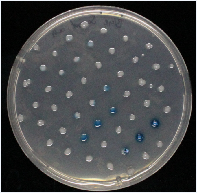
<h1><b>Beta-galactosidase</b> (LacZ)</h1> | <h1><b>Beta-galactosidase</b> (LacZ)</h1> | ||
| - | [[File:XGAL.png|left| | + | [[File:XGAL.png|left|300px|thumb|<b>Fig.5: X-Gal on LacZ expressing colony</b>]] |
| - | [[File:Green_beta_gal_on_t7_strain.JPG|left| | + | [[File:Green_beta_gal_on_t7_strain.JPG|left|300px|thumb|<b>Fig.6: Beta-green-X-gal on LacZ expressing colony</b>]] |
<br clear="all"/> | <br clear="all"/> | ||
<h1><b>Alkaline phosphatase</b> (PhoA)</h1> | <h1><b>Alkaline phosphatase</b> (PhoA)</h1> | ||
| - | [[File:PNPP_on_phoA_t7_strain.JPG|left| | + | [[File:PNPP_on_phoA_t7_strain.JPG|left|300px|thumb|<b>Fig.2: NPP on PhoA expressing colony</b>]] |
<br clear="all"/> | <br clear="all"/> | ||
<h1><b>Carboxyl esterase</b> (Aes)</h1> | <h1><b>Carboxyl esterase</b> (Aes)</h1> | ||
| - | [[File:Magenta_butyrate_on_aes_t7_strain.JPG|left| | + | [[File:Magenta_butyrate_on_aes_t7_strain.JPG|left|248px|thumb|<b>Fig.1: Magenta butyrate on Aes expressing colony</b>]] |
<br clear="all"/> | <br clear="all"/> | ||
<h1><b>Glycoside hydrolase</b> (NagZ)</h1> | <h1><b>Glycoside hydrolase</b> (NagZ)</h1> | ||
| - | [[File:XGlcNAc_on_nagZ_t7_strain.JPG|left| | + | [[File:XGlcNAc_on_nagZ_t7_strain.JPG|left|300px|thumb|<b>Fig.4: X-Glucnac on NagZ expressing colony</b>]] |
<br clear="all"/> | <br clear="all"/> | ||
<h1><b>Beta-glucuronidase</b> (GusA)</h1> | <h1><b>Beta-glucuronidase</b> (GusA)</h1> | ||
| - | [[File:Salmon_glcUA_on_gusA_t7_strain.JPG |left| | + | [[File:Salmon_glcUA_on_gusA_t7_strain.JPG |left|280px|thumb|<b>Fig.3 Magenta glucuronide on GusA expressing colony</b>]] |
<br clear="all"/> | <br clear="all"/> | ||
<br><br> | <br><br> | ||
Revision as of 11:43, 27 August 2013
Contents |
Enzyme-substrate reactions
We have cloned fluorescent receiver systems as backup for our circuit in case the hydrolase reaction do not work properly.
The enzyme substrate reactions take less than 5 minutes and are visible by eye.
Our minesweeper become better and better so keep on track for updates !
| Hydrolase | Complementary substrate / IUPAC name | Visible color | Concentration[M] | Time(s)for colorimetric response | ||
|---|---|---|---|---|---|---|
| LacZ | Beta-Galactosidase | X-Gal | 5-Bromo-4chloro-3-indolyl-beta-galactopyranoside | Blue | ||
| LacZ | Beta-Galactosidase | Green-beta-D-Gal | N-Methyl-3-indolyl-beta-D_galactopyranoside | Green | ||
| GusA | Beta-glucuronidase | Magenta glucuronide | 6-chloro-3-indolyl-beta-D-glucuronide-cycloheylammonium salt | Red | ||
| PhoA | Alkaline phosphatase | pNPP | 4-Nitrophenylphosphatedi(tris) salt | Yellow | ||
| Aes | Carboxyl esterase | Magenta butyrate | 5-bromo-6-chloro-3-indoxyl butyrate | Magenta | ||
| NagZ | Glycoside hydrolase | X-glucosaminide X-Glunac | 5-bromo-4-chloro-3-indolyl-N-acetyl-beta-D-glucosaminide | Blue | ||
What about the hydolases ? How do they work and where do they come from ? Why do we use hydrolases ?
Beta-galactosidase (LacZ)
Alkaline phosphatase (PhoA)
Carboxyl esterase (Aes)
Glycoside hydrolase (NagZ)
Beta-glucuronidase (GusA)
Do the substrates and enzymes cross-react ?
An enzyme-substrate test matrix (Figure 6) was established to test each substrate against each enzyme. The results were as expected (Figure 6.2) and no cross reaction is visible. The NagZ-X - glucosaminide X-Glunac reveal some difficulties in the liquid culture as well as on the agar plate.
 "
"


