Glucocorticoid sensor
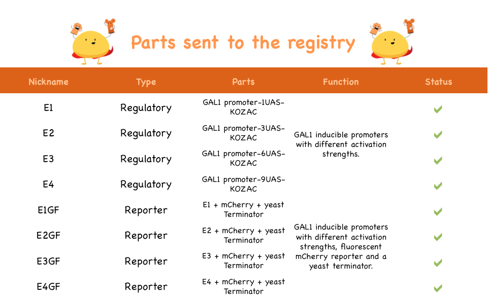
Here are the parts sent to the registry:
You can see the confirmation gels of the parts below:
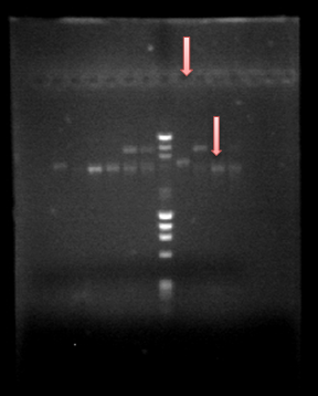
PCR confirmation: E1GF-E1GF(2)-E2GF-E2GF(2)-WM-E3GF-E3GF(2)-E4GF-E4GF(2) 
Co-transformation: WM-E1GF2-E2GF-E3GF1-E3GF2-E4GF-WM- E3GF3-E4GF2-E2GF2 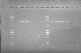
PCR confirmation of the parts cloned in the pSB1C3 backbone using the specifically designed primers 15 and 31 (see primers)
Also, the fluorescence assays for the E*GF parts

Unsaturated curve where we see how the mCherry reporter appears after the addition of 10 uM dexamethasone, a glucocorticoid. Emmision intensity was measured at 607nm after excitation at 586nm

Saturated curve for fluorescence. After three hours of the addition of 10 uM dexamethasone the reporter (mCherry) reaches its highest signal. We can see the different strengths depending on the number of UAS boxes. Emmision intensity was measured at 607nm after excitation at 586nm
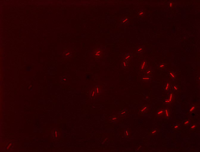
Cells with the E2GF part (BBa_K1144006) showing their fluorescent reporter, mCherry, after induction with 10 uM dexamethasone! The filter used was TRITC 
Cells with the E4GF part (BBa_K1144008) showing their fluorescent reporter, mCherry, after induction with 10 uM dexamethasone! The filter used was TRITC 2
 "
"


