Team:ETH Zurich/Experiments 3
From 2013.igem.org
Enzyme-substrate reactions
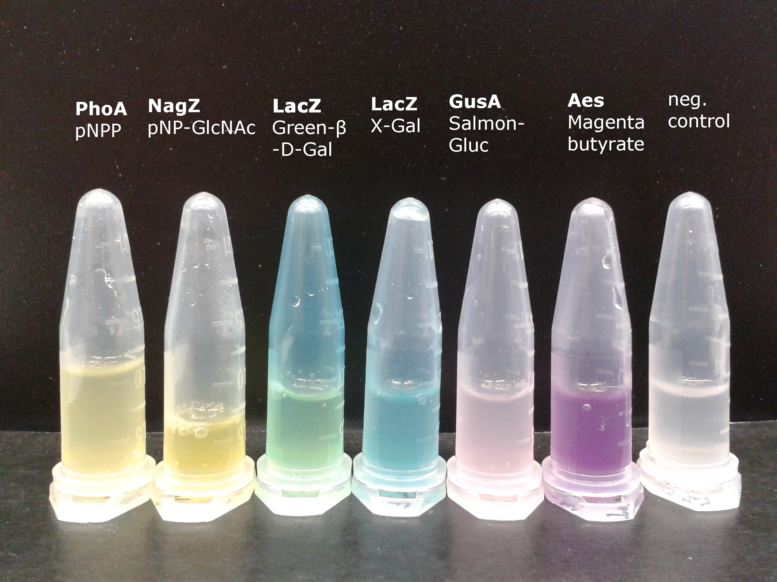
To generate visible output by adding substrates to colonies in Colisweeper, we made use of orthogonal enzyme-substrate reactions. A set of chromogenic substrates was chosen to produce different colors depending on the abundant hydrolases and thereby to uncover the identity of each colony to the player.
The set of enzyme-substrate pairs chosen for the Colisweeper project, and characterizations of their reactions, are described below. Other possible substrates that can be used for the enzymes of the Colisweeper reporter system can be found in the reporter system section.
Click on the hydrolases to see their characterization
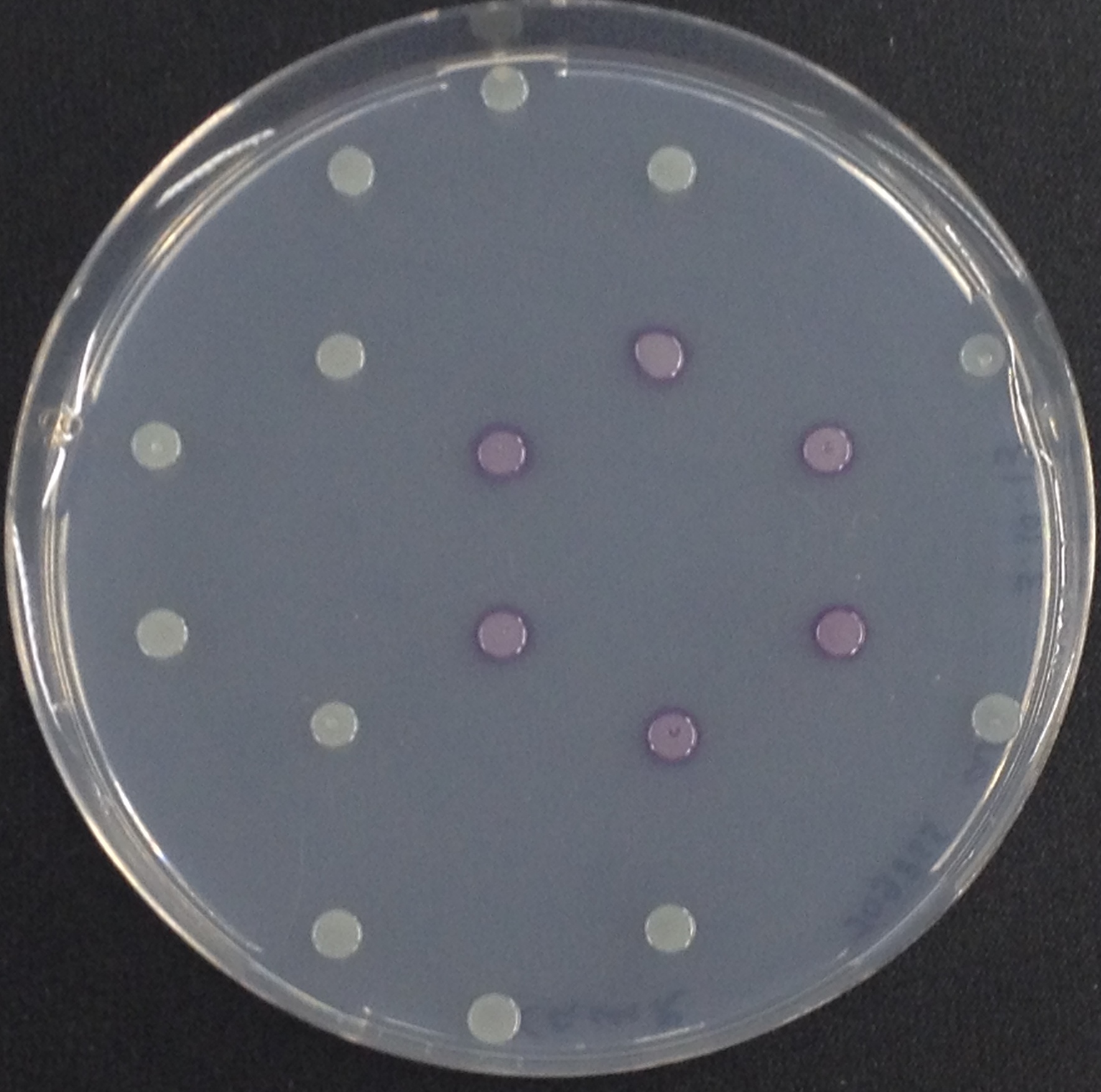
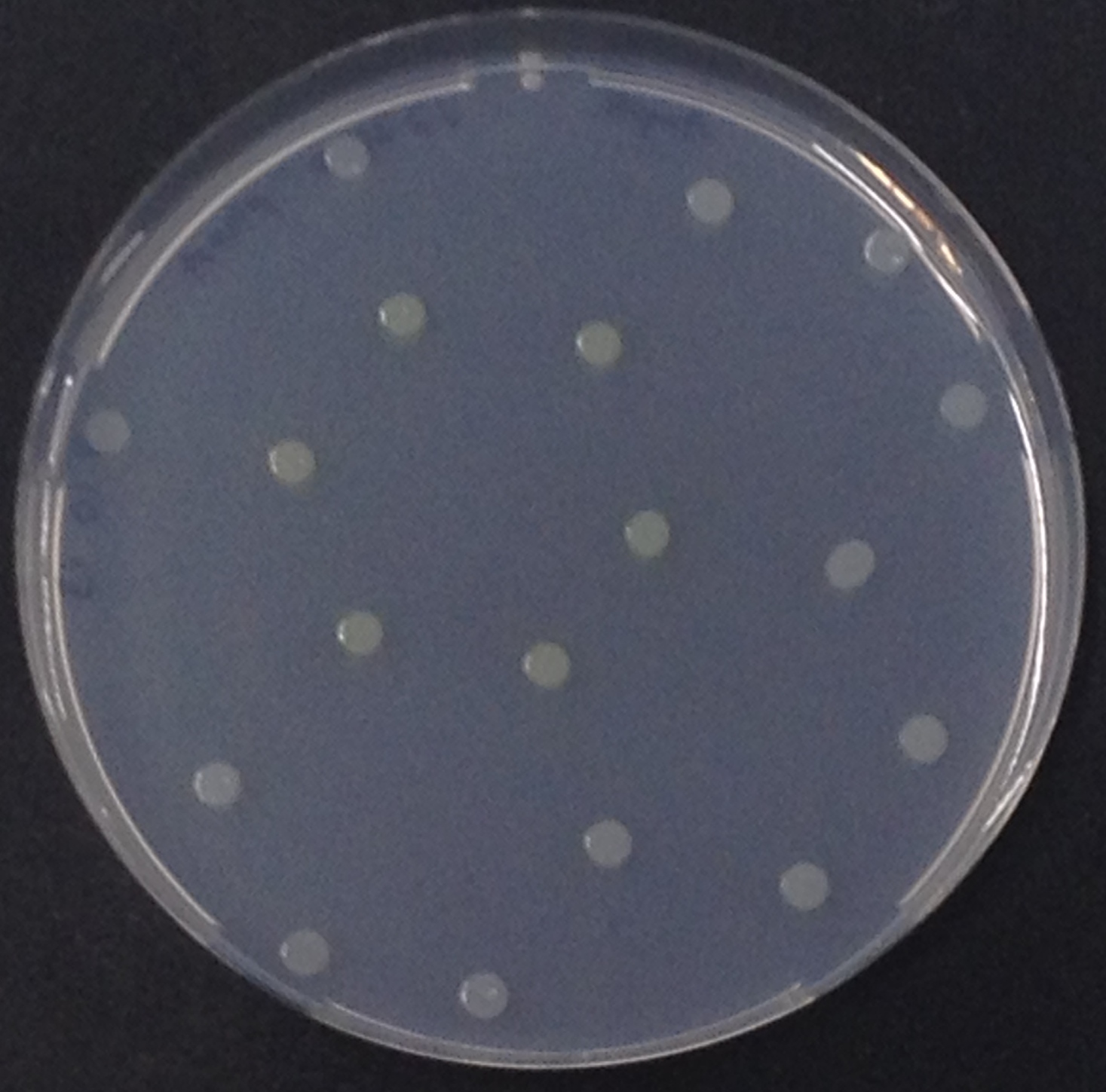
To assess color development after reaction of the enzyme with the chromogenic substrate, a liquid culture of our triple knockout Escherichia coli strain overexpressing Aes was grown until an OD600 of 0.4 - 0.6 was reached before addition of 5-Bromo-6-Chloro-3-indolyl butyrate (magenta butyrate) to a final concentration of 250 µM. To study the color development in the actual Colisweeper game setup, colonies were plated by pipetting 1.5 µl of triple knockout Escherichia coli liquid culture (OD600 of 0.4 - 0.6) on an M9 agar plate. Addition of 1.5 µl of 20 mM magenta butyrate onto each colony results in color generation visible after a few minutes at room temperature.
Enzyme kinetics
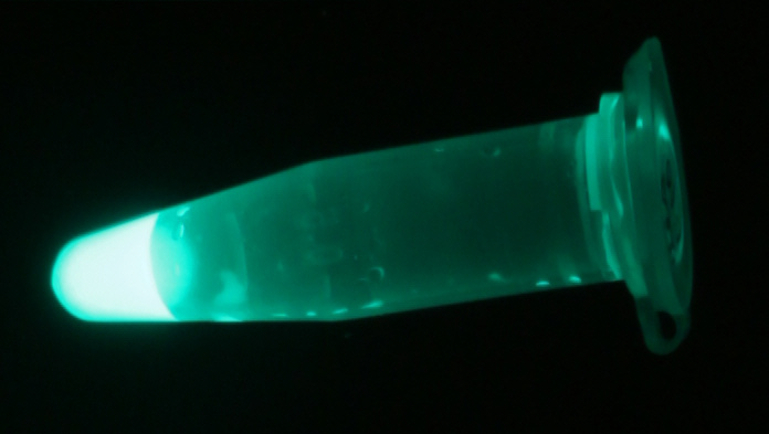
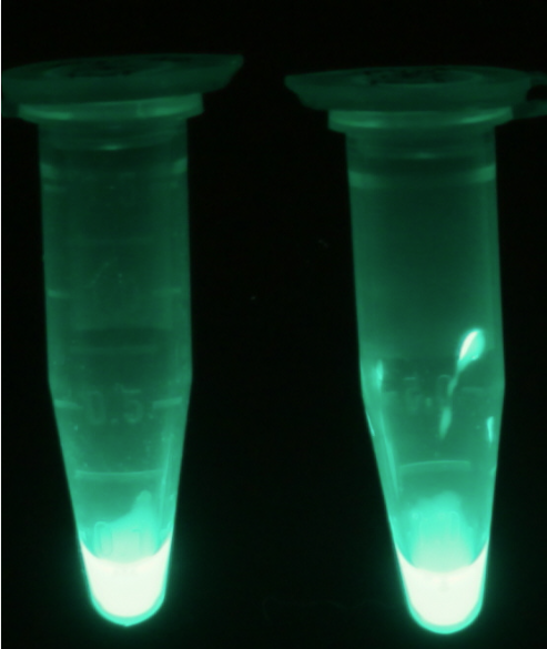
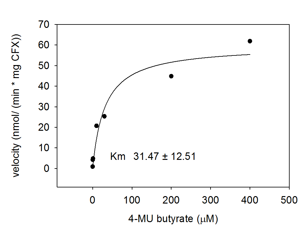
For the kinetics assay, the aes encoded protein was tested with the fluorescent substrate 4-MU-butyrate. The picture on the right was taken with a common single lens reflex camera mounted on a dark hood at λEx 365 nm.
To characterize the enzyme, we conducted fluorometric assays to obtain Km values. Bacterial cells were harvested and lysed, and the cell free extract (CFX) was then collected for the fluorometric assay. The properly diluted CFX was measured on a 96 well plate in triplicates per substrate concentration. A plate reader took measurements at λEx 365 nm and λEm 445 nm. The obtained data was evaluated and fitted to Michaelis-Menten-Kinetics with SigmaPlot™.
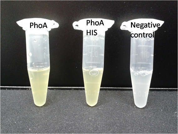
To test the functionality of this PhoA, a liquid culture of Escherichia coli overexpressing this hydrolase was grown until an OD600 of 0.4 - 0.6 was reached before addition of p-nitrophenyl phosphate (pNPP) to a final concentration of 5 µM. This substrate gives rise to yellow color after hydrolysis by the PhoA. In the Colisweeper game, substrates are pipetted onto colonies. Figure 10 shows color development on colonies five minutes after addition of 1.5 µl of a 0.5 M pNPP solution.
Characterization of PhoA-HIS tagged protein
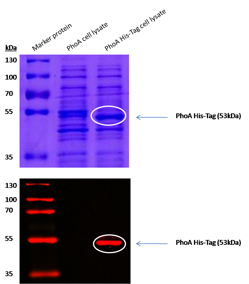
The PhoA and PhoA with HIS-tag were overexpressed in the Escherichia coli BL21 DE3 strain, induced by 5 μM IPTG (iso-propy-β-D-1-thiogalactopyranoside) and finally harvested in order to obtain the cell lysate by lysozyme lysis. This cell lysate of both cultures were analyzed next to each other in two SDS-PAGE gels, one for comassie staining (blue gel in the picture) and one for western blotting (black picture) with Anti-6X His tag® antibody from mouse and a second reporter goat anti mouse antibody with a IRDye 680RD. In the picture we can distinguish the PhoA His (53 kDa) on the western blot as well as on the SDS-PAGE gel (see white circles).
Enzyme kinetics
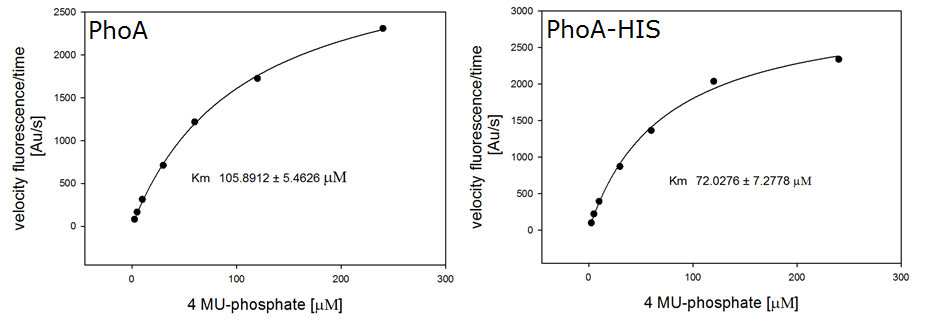
For the kinetics assay, we tested both the Citrobacter phoA and phoA-HIS encoded proteins with the fluorescent substrate 4-MU-phosphate. The picture on the right was taken with a common single lens reflex camera mounted on a dark hood at λEx 365 nm.
To characterize the enzymes, we conducted fluorometric assays to obtain Km values. Bacterial cells were harvested and lysed, and the cell free extract (CFX) was then collected for the fluorometric assay. The properly diluted CFX was measured on a 96 well plate in triplicates per substrate concentration. A plate reader took measurements at λEx 365 nm and λEm 445 nm.
The obtained data was evaluated and finally fitted to Michaelis-Menten-Kinetics with SigmaPlot™:
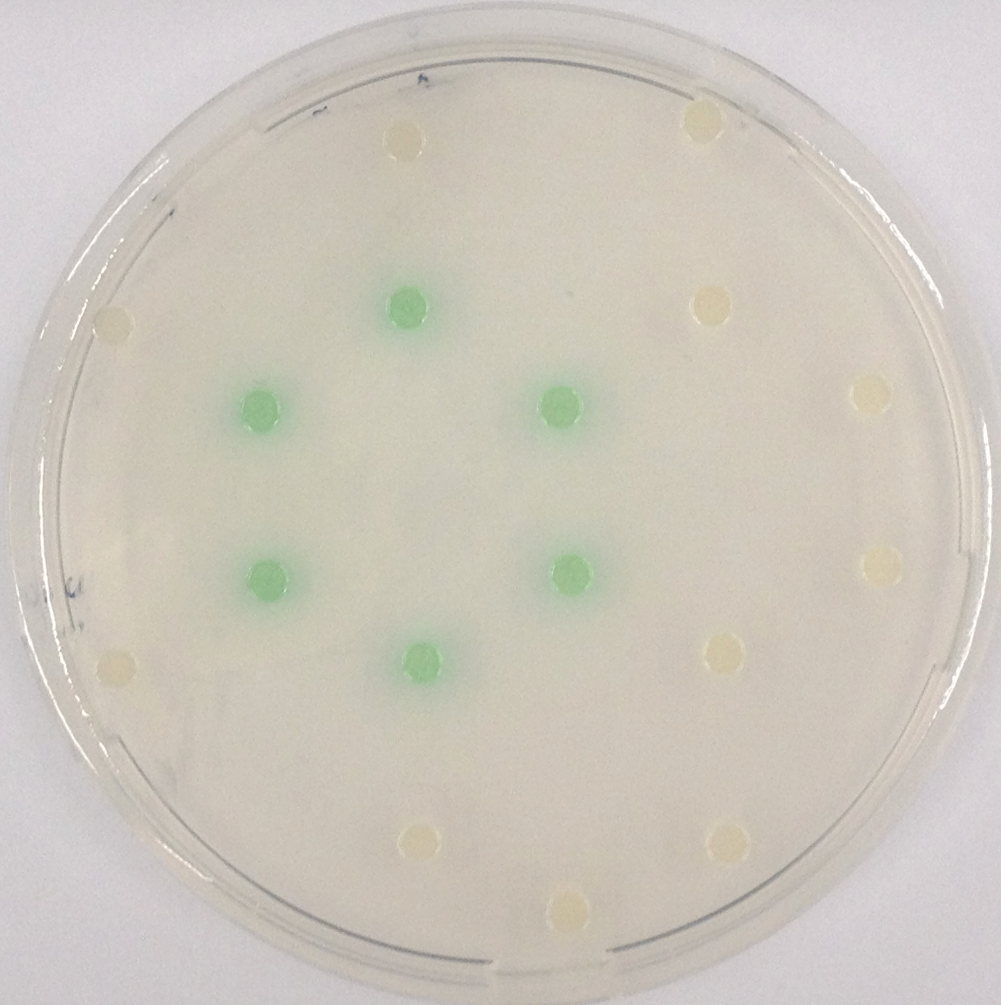
The β-Galactosidase is natively expressed in the triple knockout Escherichia coli strain used in Colisweeper. Therefore, this protein catalyzes hydrolysis of the flagging substrate, which is N-Methyl-3-indolyl-β-D-Galactopyranoside (Green-β-D-Gal) and produces a green color upon cleavage of the chromophore.
This enzyme could also be used as a mine reporter. On a separate plasmid in the mine cells under a different promoter than pLac. Growing the colonies on glucose rich medium would shut down LacZ in all other cells, rendering the mines the only ones responsive/LacZ producing.
To test the functionality of GusA, a liquid culture of Escherichia coli overexpressing GusA was grown until an OD600 of 0.4 - 0.6 was reached before addition of 6-Chloro-3-indolyl-β-glucuronide (Salmon-Gluc), a substrate for GusA which produces a rose/salmon color after cleavage. The final concentration of the substrate in liquid culture was 1 mM and the conversion of this substrate was done by overnight incubation at 37°C.
Enzyme kinetics
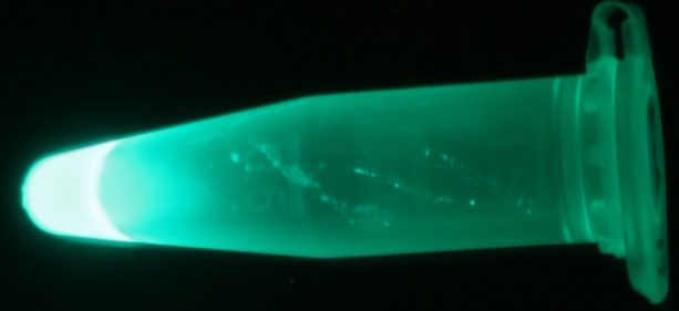
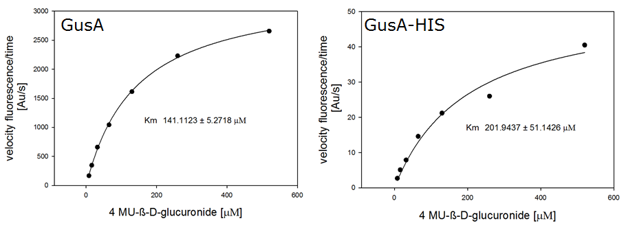
For the kinetics assay, we tested both gusA and gusA-HIS encoded proteins with the fluorescent substrate 4-MU-β-D-Glucuronide. The picture on the right was taken with a common single lens reflex camera mounted on a dark hood at λEx 365 nm.
To characterize the enzymes, we conducted fluorometric assays to obtain Km values. Bacterial cells were harvested and lysed, and the cell free extract (CFX) was then collected for the fluorometric assay. The properly diluted CFX was measured on a 96 well plate in triplicates per substrate concentration. A plate reader took measurements at λEx 365 nm and λEm 445 nm. The obtained data was evaluated and finally fitted to Michaelis-Menten-Kinetics with SigmaPlot™:
In Colisweeper, the NagZ is constitutively expressed in the mine cells. Addition of 5-Bromo-4-Chloro-3-indolyl-β-N-acetylglucosaminide (X-GluNAc) would result in a blue color precipitate, as shown in Figure 25. However, addition of the X-GluNAc substrate that we have did not result in any color production. Control assays with p-nitrophenyl-β-N-acetylglucosaminide (pNP-GluNAc) on the other hand gave rise to yellow color, which confirms that NagZ is expressed in our mine cells.
References
(1)
 "
"


